quarta-feira, 2 de julho de 2014

How do ants get around? Ultra-sensitive machines measure their every step


July 1, 2014

Society for Experimental Biology

How do ants manage to move so nimbly whilst coordinating three pairs of legs and a behind that weighs up to 60 percent of their body mass? Scientists have recently developed a device that may reveal the answer and could even help design micro-robots in the future. Researchers used an elastic polycarbonate material to produce a miniature force plate. Springs arranged at right angles to each other enabled forces to be measured across the plate in the micro-Newton range.


Red Ant (stock image). Ants walk using an "alternating tripod" system: the front and back legs of one side and the middle leg of the other side move together during one step.

How do ants manage to move so nimbly whilst coordinating three pairs of legs and a behind that weighs up to 60% of their body mass? German scientists have recently developed a device that may reveal the answer.

Measuring the forces generated by single limbs is vital to understanding the energetics of animal locomotion. However, with very small animals such as insects, this becomes problematic. Dr Reinhardt (Friedrich-Schiller University) used an elastic polycarbonate material to produce a miniature force plate. Springs arranged at right angles to each other enabled forces to be measured across the plate in the micro-Newton range.

The ants (Formica polyctena) walk using an "alternating tripod" system: the front and back legs of one side and the middle leg of the other side move together during one step. It was unknown, however, if a different gait is used for faster running speeds. Ants were made to travel down a runway built on top of the force plate, equipped with a high-speed camera to record a motion sequence. The researchers found that the basic alternating tripod gait did not alter at higher speeds, with the ants instead increasing their stride length and number of steps. The ants appear to adopt a strategy known as "grounded running"; that is, they reach higher speeds without using an "aerial phase" when all joints lose contact with the ground. This improves stability by keeping the centre of mass low, reducing the risk of falling and helping the ants to turn quickly & travel over rough terrain.

The device was also used to investigate ants travelling up a vertical surface. "During level locomotion, the typical vertical force of an ant leg is around 70 µN" Dr Reinhardt described. "The situation is different in vertical climbing. The front legs generate forces as large as the body weight -- around 20 mg. We expect that the animals can still generate much larger forces, for instance when transporting food or during fights."

The force plate was built using stereolithography technology. This uses a special photocurable polymer which solidifies when exposed to ultra-violet light. A vat is filled with the liquid polymer and a UV laser scanned across it to build up the structure layer by layer. Because the laser can be set to trace any design, this technology could be used in a wealth of applications. "Our measuring device can be applied far beyond the field of insect biomechanics" Dr Reinhardt stated. "For example, the force plate could be invaluable to the design and testing of micro-robots."


Story Source:

The above story is based on materials provided by Society for Experimental Biology. Note: Materials may be edited for content and length.

Epidemia de Ebola preocupa OMS

Ministros da Saúde de onze países da África Ocidental e especialistas internacionais abriram nesta quarta-feira uma cúpula de dois dias para examinar a opção de um "plano radical" de luta contra a epidemia mais mortal da história do vírus Ebola.

A epidemia afeta atualmente Serra Leoa, Guiné e Libéria.

Segundo o último balanço da Organização Mundial da Saúde (OMS), divulgado nesta terça-feira, os três países vizinhos somam, desde o começo do ano, 759 casos de febre hemorrágica, entre eles, 467 mortais.

São 129 casos mortais a mais que o balanço anterior - um número 38% maior -, que remonta à semana passada, um sinal de que a epidemia voltou a disparar após uma trégua no mês de abril.

"Trata-se da maior epidemia em termos de pessoas afetadas, de mortos e extensão geográfica", destacou em um comunicado a OMS, que organiza o encontro na capital de Gana.

"As decisões que forem tomadas durante esta reunião serão determinantes para combater a epidemia atual e as que estão por vir", acrescentou a organização no comunicado.

Diante do aumento contínuo da mortalidade e do número de pessoas afetadas pelo vírus Ebola, a OMS advertiu que serão "necessárias medidas drásticas" para conter esta epidemia mortal e altamente contagiosa.

Em 23 de junho, a ONG Médicos sem Fronteiras (MSF) já tinha advertido que a epidemia estava "fora de controle" e ameaçava se propagar para outras regiões.

A OMS, que enviou 150 especialistas a campo desde a primeira aparição do vírus na Guiné, em janeiro passado, compartilha desse prognóstico.

Apesar dos esforços da OMS e de outras agências especializadas, houve uma "alta importante" das taxas de novos casos e de mortes nas últimas semanas, segundo o comunicado.

Five-legged kangaroo? Telling the tale of a kangaroo's tail


Illustration of the kangaroo's fore limb, hind limb, and tail skeletal structures with corresponding photo of a red kangaroo.

Kangaroos may be nature's best hoppers. But when they are grazing on all fours, which is most of the time, their tail becomes a powerful fifth leg, says a new study.

Involving researchers at the University of Colorado Boulder, Simon Fraser University in Burnaby, Canada, and the University of New South Wales in Sydney, Australia, the study illuminates the seemingly mundane task of foraging by red kangaroos. While such activity appears awkward, it turns out their tails provide as much propulsive force as their front and hind legs combined as they eat their way across the landscape.

"We found that when a kangaroo is walking, it uses its tail just like a leg," said Associate Professor Maxwell Donelan of Simon Fraser University, corresponding author for the study. "They use it to support, propel and power their motion. In fact, they perform as much mechanical work with their tails as we do with one of our legs."

"We went into this thinking the tail was primarily used like a strut, a balancing pole, or a one-legged milking stool," said Associate Professor Rodger Kram of CU-Boulder's Department of Integrative Physiology, a study co-author. "What we didn't expect to find was how much power the tails of the kangaroos were producing. It was pretty darn surprising."

Red kangaroos are the largest of the kangaroo species in Australia. When grazing on grasses, they move both hind feet forward "paired limb" style while using their tails and front limbs together to support their bodies. "They appear to be awkward and ungainly walkers when one watches them moseying around in their mobs looking for something to eat," said Kram. "But it turns out it is not really that awkward, just weird."

In human locomotion, the back foot acts as the gas pedal and the front foot acts as a brake, which is not especially efficient, said Kram. But he likens a walking kangaroo to a skateboarder who has one foot on the board and uses the other foot -- in this case a tail -- to push backward off the pavement, increasing the forward motion.

A paper on the subject was published online in Biology Letters. In addition to Kram and Donelan, the paper was co-authored by Postdoctoral Fellow Shawn O'Connor of Simon Fraser and Emeritus Professor Terence Dawson of the University of New South Wales. The study was funded by the Natural Sciences and Engineering Research Council of Canada, the Australian Research Council, and traveling fellowships from the International Society for Biomechanics and the Journal of Experimental Biology.

Donelan, a former graduate student under Kram, said no animal other than the kangaroo uses its tail like a leg. "Their tails have more than 20 vertebrae, taking on the role of our foot, calf, and thigh bones."

The research project had its beginnings in 1973, when Dawson, a visiting professor at Harvard University, was working with Harvard Professor Richard Taylor, who later became Kram's advisor. Dawson coaxed a small group of kangaroos to hop and walk on a large motorized treadmill, with a goal of measuring the energy costs of locomotion at varying speeds. Dawson and Kram eventually showed that a kangaroo can increase its metabolism by 50 times during exercise.

"Kangaroos are really special mammals," said Dawson. "Work over the past half century has turned the notion that they belong to an inefficient, primitive group of mammals totally on its head."

The kangaroo tail also acts as a dynamic, springy counterbalance during hopping and boosts balance when male kangaroos grab each other by the chests or shoulders, then rear back and kick each other in the stomach in an attempt to assert dominance for the purpose of reproduction.

For the study the team videotaped five red kangaroos in Dawson's Sydney lab that had been trained to walk forward on a force-measuring platform with Plexiglas sides. The platform's sensors measured vertical, backward and forward forces from the legs and tails of the animals. The kangaroos had been taught that walking forward on the platform resulted in being rewarded with sweet treats, said Kram.

Over his career Kram and his students have studied the locomotion of a number of creatures, from elephants, tortoises and llamas to ostriches and beetles.

Although much of the data for the new study was collected years ago, other research efforts by the team members slowly pushed some of the key kangaroo locomotion data to the back burner. "But this was a study we just could not let go of," said Kram. "It was just too much fun. It's a real wonder of nature, how these kangaroos move about and what they are able to do."

Kram calls the evolution of the kangaroo tail, which is thought to have been prehensile when opossum-like kangaroo ancestors were living in trees, an "exaptation" -- a shift in the function of a biological trait over time. He likened it to a roll of duct tape in the back of a truck. "You know you are going to use it, you just don't know when," he said.

"I'm envious of kangaroos," said Kram, a competitive master runner in the mile and 1,500 meters. "When they hop faster, they don't use energy at a faster rate. The have the ability to move faster and not get tired, the ultimate goal of a runner."

Brazilian Obsessions: 10 Must-See Destinations in Brazil


In its young history, Brazil has established itself as one of the most exotic, culturally diverse nations in the modern world.  Its people, its nature, its cuisine and its architecture are amongst the world’s most beautiful– and we are hopelessly obsessed.  Join us to explore 10 of Brazil’s most amazing destinations, from the beaches of Ipanema to the metropolis of Sao Paulo.  Our journey starts in Rio de Janeiro, the stunning city celebrated in the video above.


Ipanema, Rio de Janeiro

In the 1960s, a cultural revival was thriving in the beach neighborhood of Ipanema.  Artists, poets and musicians would gather in Ipanema to celebrate its world-class beauty and vibrant culture.  In writing an ode to this neighborhood, composer Antonio Carlos Jobim and lyricist Vinicius de Moraes penned a song about a beautiful young girl who walked the streets of Ipanema each day on her way to the ocean.  Nearly fifty years after it was written, “Garota de Ipanema” (“The Girl from Ipanema“) remains a cultural icon of Brazil, just as the Ipanema neighborhood remains a cultural epicenter of Rio de Janeiro.  The streets of Ipanema and the beach at their end are celebrated as some of the world’s most beautiful, with the Travel Channel calling Ipanema the sexiest beach in the world.

Ipanema Gallery

Lapa, Rio de Janeiro

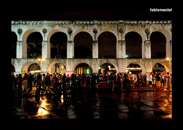
Across town from Ipanema is another cultural hotspot, the Lapa neighborhood of Rio de Janeiro.  Located in Rio’s urban center, Lapa is host to 18th century architecture, a growing arts community and an active nightlife scene.  Amongst Lapa’s most recognizable landmarks is the Arco de Lapa, a series of raised aqueducts constructed in the mid 1700s.  On Saturdays, a street party is held adjacent to a section of the Arcos in Lapa, a weekly celebration for natives and tourists alike.  Another point of interest in Lapa is the stunning-yet-strange Metropolitan Cathedral of Rio, a brutalist concrete cone that stretches 80 meters into the air.

Lapa Gallery

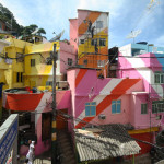
The Painted Favelas of Rio de Janeiro

Far beyond the shinier parts of Rio are the famed “favelas” of the city, or large shanty towns that are home to Rio’s poorest.  If you’re comfortable with your Portuguese and your self-defense, a guided visit to the Painted Favelas of Rio reveals a sight to behold.  A duo of artists worked with a few favelas in Rio to engender new pride in their homes.  Jeroen Koolhaas and Dre Urhahn worked with the locals to paint large sections of favelas into bright, colorful works of art.  The result was a down-on-its-luck neighborhood that had been transformed by art into a place of pride for its inhabitants.  Enter at your own risk, but be prepared for some truly inspirational, transformational art.

Favela Painting Gallery

Corcovado, Rio de Janeiro

Another historic Rio landmark is also the inspiration for another bossa nova hit.  The mountain of Corcovado rises high above the Rio de Janeiro bay with an open-armed Christ at its peak.  The famous Christ The Redeemer statue is a point of pilgrimage for Christians throughout Brazil and the world beyond, the host to weddings and other religious ceremonies held daily.  Its view of Rio is unparalleled, showing the famed Sugarloaf Mountain and Copacabana beach in the distance.  The statue above and the harbor below have each been listed as wonders of the world, and a visit to Corcovado will show precisely why.

Corcovado Gallery

The Architecture of Oscar Niemeyer

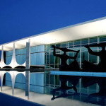
Oscar Niemeyer is arguably the greatest Brazilian architect that has ever lived, a man that gave face to many of Brazil’s most important government, museum and landmark buildings.  Niemeyer’s designs can be seen everywhere from Rio to Sao Paulo to Brasilia, the new capitol of Brazil.  In Brasilia alone, Niemeyer designed the Palácio da Alvorada (the home of Brazil’s presidents), the National Congress Building, the Supreme Court, the National Theater, the Ministry of Justice, the National Library and more.  Niemeyer championed the contemporary movement in Brazil, giving a bold character to a government that serves a bold, proud people.  Niemeyer’s buildings should be visited in Brazil, and you’ll find them in nearly every major city you might visit.  Keep your eye out for Oscar himself, as the architect is still at work today– at the bright young age of 103 years old.  (see more Brazilian Architecture including Niemeyer designs here on TheCoolist)

Oscar Niemeyer Gallery

Paraty, RJ, Brazil

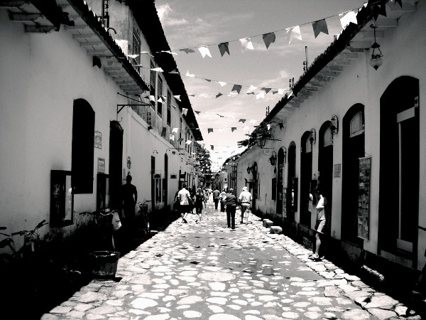
While Rio de Janeiro is deserving of a long vacation of its own, there is so much more to see in Brazil at large.  A short trip down the coast from Rio is Paraty, Brazil, a quiet seaside town that serves up a slower pace of life and a stunning natural environment.  Paraty was founded by Portuguese settlers in the late 1600s, built primarily on the rise of the Brazilian gold rush.  Paraty’s architecture recalls the Portuguese style of the 17th/18th centuries, but its coast has become more progressive in recent years.  Our favorite work of residential architecture in this young century is located in Paraty, the Paraty House by Brazilian architect Marcio Kogan and company.  If it were up to us, we’d do just about anything for a day in the quiet streets of Paraty and a night in that home by Marcio Kogan.

Paraty Gallery

Iguazu Falls

One of the world’s most massive and visually stunning waterfalls is claimed by both Brazil and Argentina, a section of water that is also a national border.  Iguazu Falls are amongst the widest in the world, displacing up to 12,000 cubic meters of water per second– the fastest rate of any waterfall on Earth.  The Iguazu Falls are split into over 250 individual waterfalls with terraces and islands between them.  Visitors to Iguazu can stand at a central point between the waterfalls to be surrounded by the falls in 260 degrees.  While other falls may be bigger or have a more continuous curtain of water, it is hard to argue that any others are as beautiful, as the rare formation of land and cliffs provides vistas that other waterfalls cannot claim.

Iguazu Falls Gallery

Ouro Preto, MG, Brazil

Ouro Preto is located inland in the great state of Minas Gerais, a mining town with great cultural history.  Ouro Preto is one of Brazil’s many UNESCO World Heritage sites, known for its early Baroque architecture and other inspirations from its European forefathers.  Ouro Preto hosts plenty of respected museums, tour-friendly mines and fantastic churches, enough that Ouro Preto is amongst Brazil’s top tourist destinations.  The whole of Minas Gerais is dotted with many towns like Ouro Preto with rich cultural history, many of them born from the gold boom of the late 17th century.

Ouro Preto Gallery

Sao Paulo, Brazil

With a population of over 11,000,000 people, Sao Paulo is by far the largest city in the Americas.  It’s the largest in both the Southern and Western Hemispheres, and its public transit system is the world’s busiest.  Sao Paulo may not have a travel appeal as recognizable as Rio, but it is a city that must be experienced by every self-respecting world traveler.  Its mix of cultures have yielded a brilliant culinary, art and music scene and a wild architectural identity.  Its size is truly staggering, and while this great city deserves a destination list of its own, a traveler through Brazil is required to experience it.

Sao Paulo Gallery

Salvador, BA, Brazil

Salvador,BA,is the capital of Bahia, the final stop on our tour of Brazil, a city known as Brazil’s “capital of happiness”.  Salvador is one of the oldest cities in Brazil and the Americas in general, dating back to 1549.  Salvador is largely an African American city, with over 80% of its population of African descent.  As a result, Salvador has rich African cultural influences alive in its cuisine, its dance and its artistry.  Brazil’s famous Capoeira, a mix of dance and martial artistry, was born in Salvador from early Afro-Brazilian performers.  Salvador is also home to the largest street party on the planet, the world’s greatest Carnaval party with nearly 2,000,000 in attendance.

Salvador, BA, Brazil

Got the itch?  A trip to Brazil should most certainly be on your itinerary.  This culturally vibrant country is home to beautiful people, rich cuisine, old world architecture and stunning vistas of environmental paradise.  If you have a story or a tip about visiting Brazil, please do share it with us in the comments.  These are 10 of our favorite destinations, we’d love to hear about the great places that we missed.

In human evolution, changes in skin's barrier set northern Europeans apart


June 30, 2014

University of California, San Francisco (UCSF)

The popular idea that northern Europeans developed light skin to absorb more UV light so they could make more vitamin D -- vital for healthy bones and immune function -- is questioned by researchers in a new study. Ramping up the skin’s capacity to capture UV light to make vitamin D is indeed important, however, researchers concluded in their study that changes in the skin’s function as a barrier to the elements made a greater contribution than alterations in skin pigment in the ability of northern Europeans to make vitamin D.


The Elias lab has shown that pigmented skin provides a better skin barrier, which was critically important for protection against dehydration and infections among ancestral humans living in sub-Saharan Africa. But the need for pigment to provide this extra protection waned as modern human populations migrated northward over the past 60,000 years or so, while the need to absorb UVB light became greater, particularly for those humans who migrated to the far North behind retreating glaciers less than 10,000 years ago.

[show more]

The popular idea that Northern Europeans developed light skin to absorb more UV light so they could make more vitamin D – vital for healthy bones and immune function – is questioned by UC San Francisco researchers in a new study published online in the journal Evolutionary Biology.

Ramping up the skin’s capacity to capture UV light to make vitamin D is indeed important, according to a team led by Peter Elias, MD, a UCSF professor of dermatology. However, Elias and colleagues concluded in their study that changes in the skin’s function as a barrier to the elements made a greater contribution than alterations in skin pigment in the ability of Northern Europeans to make vitamin D.

Elias’ team concluded that genetic mutations compromising the skin’s ability to serve as a barrier allowed fair-skinned Northern Europeans to populate latitudes where too little ultraviolet B (UVB) light for vitamin D production penetrates the atmosphere.

Among scientists studying human evolution, it has been almost universally assumed that the need to make more vitamin D at Northern latitudes drove genetic mutations that reduce production of the pigment melanin, the main determinant of skin tone, according to Elias.

“At the higher latitudes of Great Britain, Scandinavia and the Baltic States, as well as Northern Germany and France, very little UVB light reaches the Earth, and it’s the key wavelength required by the skin for vitamin D generation,” Elias said.

“While is seems logical that the loss of the pigment melanin would serve as a compensatory mechanism, allowing for more irradiation of the skin surface and therefore more vitamin D production, this hypothesis is flawed for many reasons,” he continued. “For example, recent studies show that dark-skinned humans make vitamin D after sun exposure as efficiently as lightly-pigmented humans, and osteoporosis – which can be a sign of vitamin D deficiency – is less common, rather than more common, in darkly-pigmented humans.”

Furthermore, evidence for a south to north gradient in the prevalence of melanin mutations is weaker than for this alternative explanation explored by Elias and colleagues.

In earlier research, Elias began studying the role of skin as a barrier to water loss. He recently has focused on a specific skin-barrier protein called filaggrin, which is broken down into a molecule called urocanic acid – the most potent absorber of UVB light in the skin, according to Elias. “It’s certainly more important than melanin in lightly-pigmented skin,” he said.

In their new study, the researchers identified a strikingly higher prevalence of inborn mutations in the filaggrin gene among Northern European populations. Up to 10 percent of normal individuals carried mutations in the filaggrin gene in these northern nations, in contrast to much lower mutation rates in southern European, Asian and African populations.

Moreover, higher filaggrin mutation rates, which result in a loss of urocanic acid, correlated with higher vitamin D levels in the blood. Latitude-dependent variations in melanin genes are not similarly associated with vitamin D levels, according to Elias. This evidence suggests that changes in the skin barrier played a role in Northern European’s evolutionary adaptation to Northern latitudes, the study concluded.

Yet, there was an evolutionary tradeoff for these barrier-weakening filaggrin mutations, Elias said. Mutation bearers have a tendency for very dry skin, and are vulnerable to atopic dermatitis, asthma and food allergies. But these diseases have appeared only recently, and did not become a problem until humans began to live in densely populated urban environments, Elias said.

The Elias lab has shown that pigmented skin provides a better skin barrier, which he says was critically important for protection against dehydration and infections among ancestral humans living in sub-Saharan Africa. But the need for pigment to provide this extra protection waned as modern human populations migrated northward over the past 60,000 years or so, Elias said, while the need to absorb UVB light became greater, particularly for those humans who migrated to the far North behind retreating glaciers less than 10,000 years ago.

The data from the new study do not explain why Northern Europeans lost melanin. If the need to make more vitamin D did not drive pigment loss, what did? Elias speculates that, “Once human populations migrated northward, away from the tropical onslaught of UVB, pigment was gradually lost in service of metabolic conservation. The body will not waste precious energy and proteins to make proteins that it no longer needs.”

For the Evolutionary Biology study, labeled a “synthesis paper” by the journal, Elias and co-author Jacob P. Thyssen, MD, a professor at the University of Copenhagen, mapped the mutation data and measured the correlations with blood levels of vitamin D. Labs throughout the world identified the mutations. Daniel Bikle, MD, PhD, a UCSF professor of medicine, provided expertise on vitamin D metabolism.


Story Source:

The above story is based on materials provided by University of California, San Francisco (UCSF). The original article was written by Jeffrey Norris. Note: Materials may be edited for content and length.


Journal Reference:

  1. Jacob P. Thyssen, Daniel D. Bikle, Peter M. Elias. Evidence That Loss-of-Function Filaggrin Gene Mutations Evolved in Northern Europeans to Favor Intracutaneous Vitamin D3 Production. Evolutionary Biology, 2014; DOI: 10.1007/s11692-014-9282-7

Study helps unlock mystery of high-temp superconductors


A Binghamton University physicist and his colleagues say they have unlocked one key mystery surrounding high-temperature superconductivity.

A Binghamton University physicist and his colleagues say they have unlocked one key mystery surrounding high-temperature superconductivity. Their research, published this week in the Proceedings of the National Academy of Sciences, found a remarkable phenomenon in copper-oxide (cuprate) high-temperature superconductors.

Michael Lawler, assistant professor of physics at Binghamton, is part of an international team of physicists with an ongoing interest in the mysterious pseudogap phase, the phase situated between insulating and superconducting phases in the cuprate phase diagram.

“Evidence has been accumulating that this phase supports an exotic density wave state that may be key to its existence,” the physicists write in the new journal article. A density wave forms in a metal if the fluid electrons themselves crystalize.

Using a scanning tunneling microscope (STM) to visualize the electronic structure of the oxygen sites within a superconductor, the team found a density wave with a d-orbital structure. (The electron density near each copper atom looks a bit like a daisy in the crystallized pattern.) That’s especially surprising because most density waves have an s-orbital structure; their electron density is isotropic. “It’s not the pattern you would expect,” Lawler says.

In this research, Lawler and his colleagues focused on a member of the cuprate class of superconductors called bismuth strontium calcium copper oxide (BSCCO). “We now believe these density waves exist in all cuprates,” says Lawler, a theorist whose contribution to the research involved subtle uses of the Fourier transform, a mathematical analysis that’s useful when examining amplitude patterns in waves.

Superconductors conduct electricity without resistance below a certain temperature. For decades, it was thought that these materials could conduct electricity only at temperatures far below freezing. Since 1987, however, scientists have discovered several compounds that superconduct at much higher temperatures.

Development of this technology could lead to near lossless delivery of electricity to homes and businesses as well as to improvements in cell phone tower receptions and even high-speed trains.


Story Source:

The above story is based on materials provided by Binghamton University, State University of New York. Note: Materials may be edited for content and length.

'Molecular movies' will enable extraordinary gains in bioimaging, health research


Fluorescent protein biosensors provide a technology to capture the biochemical process of life almost like a motion picture that could be viewed a frame at a time. This may allow the targeted design of next-generation biosensors to track life processes and battle diseases.

Researchers have announced the creation of an imaging technology more powerful than anything that has existed before, and is fast enough to observe life processes as they actually happen at the molecular level.

Chemical and biological actions can now be measured as they are occurring or, in old-fashioned movie parlance, one frame at a time. This will allow creation of improved biosensors to study everything from nerve impulses to cancer metastasis as it occurs.

The measurements, created by the use of short pulse lasers and bioluminescent proteins, are made in femtoseconds, which is one millionth of one billionth of a second. A femtosecond, compared to one second, is about the same as one second compared to 32 million years.

That's a pretty fast shutter speed, and it should change the way biological research and physical chemistry are being done, scientists say.

Findings on the new technology were published in Proceedings of the National Academy of Sciences, by researchers from Oregon State University and the University of Alberta.

"With this technology we're going to be able to slow down the observation of living processes and understand the exact sequences of biochemical reactions," said Chong Fang, an assistant professor of chemistry in the OSU College of Science, and lead author on the research.

"We believe this is the first time ever that you can really see chemistry in action inside a biosensor," he said. "This is a much more powerful tool to study, understand and tune biological processes."

The system uses advanced pulse laser technology that is, in itself, fairly new, and builds upon the use of "green fluorescent proteins" that are extremely popular in bioimaging and biomedicine. These remarkable proteins glow when light is shined upon them. Their discovery in 1962, and the applications that followed were the basis for a Nobel Prize in 2008.

Existing biosensor systems, however, are created largely by random chance or trial and error. By comparison, the speed of the new approach will allow scientists to "see" what is happening at the molecular level and create whatever kind of sensor they want by rational design. This will improve the study of everything from cell metabolism to nerve impulses, how a flu virus infects a person, or how a malignant tumor spreads.

"For decades, to create the sensors we have now, people have been largely shooting in the dark," Fang said. "This is a fundamental breakthrough in how to create biosensors for medical research from the bottom up. It's like daylight has finally come."

The technology, for instance, can follow the proton transfer associated with the movement of calcium ions -- one of the most basic aspects of almost all living systems, and also one of the fastest. This movement of protons is integral to everything from respiration to cell metabolism and even plant photosynthesis. Scientists will now be able to identify what is going on, one step at a time, and then use that knowledge to create customized biosensors for improved imaging of life processes.

"If you think of this in photographic terms," Fang said, "we now have a camera fast enough to capture the molecular dance of life. We're making molecular movies. And with this, we're going to be able to create sensors that answer some important, new questions in biophysics, biochemistry, materials science and biomedical problems."


Story Source:

The above story is based on materials provided by Oregon State University. Note: Materials may be edited for content and length.

3-D printed wrist splints for arthritis sufferers


A Loughborough University lecturer has developed a computer software concept that will enable clinicians with no experience in Computer Aided Design (CAD) to design and make custom-made 3D printed wrist splints for rheumatoid arthritis sufferers.

A Loughborough University lecturer has developed a computer software concept that will enable clinicians with no experience in Computer Aided Design (CAD) to design and make custom-made 3D printed wrist splints for rheumatoid arthritis sufferers.

Dr Abby Paterson, from the Design School, said: "I wanted to give clinicians the ability to make splints they have not been able to make before. They can improve the aesthetics, the fit, and integrate extra bits of functionality they couldn't do before as a result of our Additive Manufacturing facilities here at Loughborough University. Thanks to our Objet Connex machine, we can integrate multiple materials in a single splint such as rubber-like integral hinges or cushioning features but, more importantly, the specialised software prototype we've developed will enable clinicians to design these splints for their patients."

The 3D printed splints are not only more comfortable and attractive but potentially cheaper than the current ones that are 'ugly, bulky, and can make a patients arm sweat'. As a result patients do not use them as often as they should.

The splints, which provide joint protection, rest, and promote pain relief,could be a major boost for sufferers of rheumatoid arthritis, the second most common type of arthritis in the UK which affects more than 400,000 people.

The splints are made by scanning a patient's arm in the 'appropriate position'. A 3D model splint is then designed based on the scan to generate a computer model.

The 3D printer can then produce as many splints as are needed at the touch of a button. They can be any colour, feature multiple materials, have a lattice design to aid ventilation and any type of fastening the patient requires.

The 3D CAD software prototype was shown to certified splinting practitioners, such as occupational therapists and physiotherapists.

Dr Paterson said: "The practitioners were very excited by new, novel ideas to expand the possibilities available to them, such as integrated rubber borders for increased comfort."

The 3D CAD software prototype is the product of Dr Paterson's PhD and development ‎work is still needed on the software and materials. ‎

Dr Paterson was supervised during her PhD by Dr Richard Bibb and Dr Ian Campbell. Dr Bibb came up with the idea for bespoke wrist splints in the late 1990's.

Dr Bibb and Dr Paterson are currently pursuing opportunities to perform a 'thorough cost analysis' of providing the service.

Dr Bibb says the 3D splints could be cheaper than the current ones because the design and manufacture stages have been separated. He believes they will be cost-effective for the NHS while the 'sky's the limit' in the private sector.

Dr Bibb, Reader in Medical Applications of Design in the Design School, said: "We are in the development phase. The research has proved that this is desirable and the clinicians want it. We know there's lots of potential."


Story Source:

The above story is based on materials provided by University of Loughborough. Note: Materials may be edited for content and length.
Biology labs: Managing the data jungle


Many biology labs fight with a glut of measurement data. New software aims to make this a thing of the past: it simplifies laboratory experiment evaluation and unifies how data is saved. It even identifies measurement errors on the spot.

During laboratory testing, countless measurement results accrue. To completely and systematically archive this body of data is extremely time consuming. In fact, researchers in the life sciences spend a quarter of their time managing data, according to an online survey of 70 people working in biology laboratories conducted by the Fraunhofer Institute for Applied Information Technology FIT in Sankt Augustin. Many of those surveyed reported that they have no centralized or structured approach to data collection in their workplace. And when a PhD student or assistant with years of experience leaves the institute and the successor tries to find and make sense of previous results, the search often begins with cryptic Excel tables and stacks of paper.

The FIT has taken steps to alleviate this problem. With its step-by-step operation, its "MPlexAnalyzer" software makes it considerably easier to manage data. Initially, the FIT experts concentrated on cytometric devices, which enable the simultaneous determination of a variety of proteins in a test batch. This approach, also called a cytometric multiplex assay, is a standard method in every biology lab. However, assays such as these are very complex and produce huge amounts of data, so it is no wonder that personnel without access to any supporting software often lose the upper hand with data collection. The FIT software's wizard assistant guides users through the entire measurement process, starting with the selection of the microtiter plates, the choice of samples, and the assignment of standard samples, and provides a clearly arranged PDF report for print out; the process is transparent and easy for beginners to grasp quickly.

A cytometric multiplex assay uses polystyrene beads with a diameter of only six micrometers. These are infused with a dye mixture that glows when subjected to two laser beams. The glowing pattern is like a fingerprint; up to a hundred different types of beads can be differentiated in this way. During the measurement, the beads flow through a thin glass cannula like pearls strung on a necklace. Cameras measure the color patterns, counting and sorting them as they go past. But biologists are primarily interested in the beads' second cargo: color-coded antibodies on the surface of each bead that are stimulated by one of the two lasers. This dye emits light at another wavelength -- but only if specific substances have bound with the antibodies, such as blood components, cell excretions or cancer cell signal proteins. A multiplex assay examines up to 100 of these substances simultaneously.

The result is a huge data set that records the number of registered beads together with the identified substance. Measurements are highly automated -- every minute, up to 96 different samples are tested, each in a little well on a glass plate. One portion of each plate is covered with samples, and another portion with reference substances used to calibrate the readings. Until now, the process of documenting where each sample is located and which measurement is recorded was a time-consuming manual task. "Our software wizard simplifies the process. With just a few mouse clicks, you can mark on the screen which wells contain reference substances or which ones are empty. If the corresponding field is red, then the specification and the measurement don't match. This means the lab assistant will see immediately if a mistake has been made or if the quality of the measurement is insufficient to reach a statistically reliable conclusion," explains Dr. Andreas Pippow, a scientist at the FIT.

For the FIT, the software is a way in to the data management market for biological labs. The idea will now be carried over to other applications, such as microscopes. At the biology lab of Fraunhofer FIT's Life Science Informatics department in Sankt Augustin, scientists are building special microscopes that can automatically shuttle large samples back and forth under the objective and scan them. There are plans for a database of all lab-performed measurements, such as those from multiplex assays, microscope images or other measuring equipment. The appeal of this common data management approach lies in the built-in ability to perform cross checks. For instance, when body cells emit certain chemical messengers as a result of a disease, often these will have consequences for the tissue structure. But the only way to spot this is by using the software to match the signal substance tested in the multiplex assay against microscope images.


Story Source:

The above story is based on materials provided by Fraunhofer-Institut fuer Angewandte Informationstechnik (FIT). Note: Materials may be edited for content and length.

Scientists discover how 'plastic' solar panels work


Three laser beams are needed to record the excited vibrational modes of PCDTBT with the method called femtosecond stimulated Raman spectroscopy. First, the green pulse is absorbed by the polymer, just as sunlight would be in a solar cell, which creates the excited state. Then, a pair of infra-red and white pulses probe this excited vibrational mode. Very short pulses of light and precise timing enable an impressive time resolution of less than 300 femtoseconds.

Scientists don't fully understand how 'plastic' solar panels work, which complicates the improvement of their cost efficiency, thereby blocking the wider use of the technology. However, researchers at the University of Montreal, the Science and Technology Facilities Council, Imperial College London and the University of Cyprus have determined how light beams excite the chemicals in solar panels, enabling them to produce charge. "Our findings are of key importance for a fundamental mechanistic understanding, with molecular detail, of all solar conversion systems -- we have made great progress towards reaching a 'holy grail' that has been actively sought for several decades," said the study's first author, Françoise Provencher of the University of Montreal.

The findings were published today in Nature Communications.

The researchers have been investigating the fundamental beginnings of the reactions that take place that underpin solar energy conversion devices, studying the new brand of photovoltaic diodes that are based on blends of polymeric semiconductors and fullerene derivatives. Polymers are large molecules made up of many smaller molecules of the same kind -- consisting of so-called 'organic' building blocks because they are composed of atoms that also compose molecules for life (carbon, nitrogen, sulphur). A fullerene is a molecule in the shape of a football, made of carbon. "In these and other devices, the absorption of light fuels the formation of an electron and a positive charged species. To ultimately provide electricity, these two attractive species must separate and the electron must move away. If the electron is not able to move away fast enough then the positive and negative charges simple recombine and effectively nothing changes. The overall efficiency of solar devices compares how much recombines and how much separates," explained Sophia Hayes of the University of Cyprus, last author of the study.

Two major findings resulted from the team's work. "We used femtosecond stimulated Raman spectroscopy," explained Tony Parker of the Science and Technology Facilities Council's Central Laser Facility. "Femtosecond stimulated Raman spectroscopy is an advanced ultrafast laser technique that provides details on how chemical bonds change during extremely fast chemical reactions. The laser provides information on the vibration of the molecules as they interact with the pulses of laser light." Extremely complicated calculations on these vibrations enabled the scientists to ascertain how the molecules were evolving. Firstly, they found that after the electron moves away from the positive centre, the rapid molecular rearrangement must be prompt and resemble the final products within around 300 femtoseconds (0.0000000000003 s). A femtosecond is a quadrillionth of a second -- a femtosecond is to a second as a second is to 3.7 million years. This promptness and speed enhances and helps maintain charge separation.

Secondly, the researchers noted that any ongoing relaxation and molecular reorganisation processes following this initial charge separation, as visualised using the FSRS method, should be extremely small. "Our findings open avenues for future research into understanding the differences between material systems that actually produce efficient solar cells and systems that should as efficient but in fact do not perform as well. A greater understanding of what works and what doesn't will obviously enable better solar panels to be designed in the future," said the University of Montreal's Carlos Silva, who was senior author of the study.


Story Source:

The above story is based on materials provided by University of Montreal. Note: Materials may be edited for content and length.

Journal Reference:

  1. Françoise Provencher, Nicolas Bérubé, Anthony W. Parker, Gregory M. Greetham, Michael Towrie, Christoph Hellmann, Michel Côté, Natalie Stingelin, Carlos Silva, Sophia C. Hayes. Direct observation of ultrafast long-range charge separation at polymer–fullerene heterojunctions. Nature Communications, 2014; 5 DOI: 10.1038/ncomms5288

Weave a cell phone into your shirt? Engineers envision an electronic switch just three atoms thick


July 1, 2014

Stanford School of Engineering

Researchers believe they've discovered a crystal that can form a monolayer three atoms thick. Computer simulations show that this crystal, molybdenum ditelluride, can act like a switch: its crystal lattice can be mechanically pulled and pushed, back and forth, between two different atomic structures -- one that conducts electricity well, the other that does not. The team hopes experimental scientists will make this semiconductor crystal and use it to fashion flexible electronics.


In the top panel, this three-atom thick crystal is shown as semiconductor that is non-conductive. An outward tug on the material (shown in the middle panel) clicks the crystal into a metallic, or conductive state. The third panel shows the crystal back in a non-conductive state.

Computer simulation shows how to make a crystal that would toggle like a light switch between conductive and non-conductive structures; this could lead to flexible electronic materials and enable a cell phone to be woven into a shirt.

Do not fold, spindle or mutilate. Those instructions were once printed on punch cards that fed data to mainframe computers. Today's smart phones process more data, but they still weren't built for being shoved into back pockets.

In the quest to build gadgets that can survive such abuse, engineers have been testing electronic systems based on a new materials that are both flexible and switchable -- that is, capable of toggling between two electrical states, on-off, one-zero, the binary commands that can program all things digital.

Now three Stanford researchers believe that they've discovered just such a flexible, switchable material. It is a crystal that can form a paper-like sheet just three atoms thick. Computer simulations show that this crystalline lattice has the remarkable ability to behave like a switch: it can be mechanically pulled and pushed, back and forth, between two different atomic structures -- one that conducts electricity well, the other that does not.

"Think of it like flicking a light switch on and off," says Karel-Alexander Duerloo, a Stanford Engineering graduate student and first author of an article in Nature Communications.

So far this discovery only exists as a simulation. But co-author and team leader Evan Reed, Assistant Professor of Materials Science and Engineering, hopes this work will inspire experimental scientists to fabricate this super-thin crystal and use it to create electronic and other devices that would be as light and flexible as fibers.

Theoretically, such electronic materials have potential to reduce battery-draining power consumption in existing devices such as smart phones. This new, power-efficient material could also make it possible to create 'smart' clothing -- imagine an ultralight cell phone or a GPS system integrated into your shirt.

Duerloo said this switchable material is formed when one atomic layer of molybdenum atoms gets sandwiched between two atomic layers of tellurium atoms.

Molybdenum and tellurium are elements that are currently used as additives for making alloys, such as steel. Tellurium is also an important component of many modern solar cells.

In his simulation, Duerloo relied on the fact that molybdenum and tellurium form a sheet-like crystal lattice that is just three-atoms thick. Notably, this atomic sandwich can form different crystalline structures that have useful properties: in one structure this lattice easily conducts electricity; in the other configuration it does not.

Duerloo's simulations show that it takes just a tiny effort to toggle the atomic structure of this three-layer amalgam from a non-conductive state into a conductive state. A gentle push switches the material back to the off state.

These simulations, as yet unsupported by experimental confirmation, are at the leading edge of a new branch of materials science that delves into the behavior of monolayer substances.

The first and most famous monolayer is graphene, which was first observed in 2004. Graphene is a layer of carbon atoms that form a lattice that resembles chicken wire. Although it is just one atom thick, graphene is incredibly strong. A sheet of graphene could bear the weight of a cat without breaking this atomically thin lattice.

Graphene is also electrically conductive. That makes it potentially useful as a light, low power electronic component.

The discoverers of graphene shared a Nobel Prize in 2010, but even before this their work was so honored that other scientists had started looking for other monolayer materials with this interesting confluence of properties: strong, stable, crystalline structures that could conduct electricity.

To help find the most promising materials from a vast universe of molecular structures, a new discipline is rising: computational materials science.

"We're like the advance scouts that survey the terrain and look for the best materials," Reed said.

Now that they have simulated the potential of this molybdenum-tellurium crystal the Stanford researchers -- the third team member is graduate student Yao Li -- hope experimental scientists will explore possible uses of this three-atom thick switch.

"No would have known this was possible before because they didn't know where to look," Duerloo said.


Story Source:

The above story is based on materials provided by Stanford School of Engineering. The original article was written by Tom Abate. Note: Materials may be edited for content and length.


Journal Reference:

  1. Karel-Alexander N. Duerloo, Yao Li, Evan J. Reed. Structural phase transitions in two-dimensional Mo- and W-dichalcogenide monolayers. Nature Communications, 2014; 5 DOI: 10.1038/ncomms5214