sexta-feira, 28 de agosto de 2015

Pregnant? Get Tdap in Your Third Trimester


Man and woman's hands on pregnant woman's stomach making shape of heart

Only you can give your baby protection against whooping cough before your little one is even born. Talk to your doctor or midwife about getting the Tdap vaccine during your third trimester.

Whooping cough is a serious disease that can be deadly for babies. Unfortunately, babies can't get vaccinated and start building protection against whooping cough until they are two months old. The good news is that you can avoid this gap in protection by getting the whooping cough vaccine (called Tdap) during the third trimester of your pregnancy. By doing so, you pass antibodies to your baby before birth. These antibodies help protect your baby in the first few months of life.

You Need a Whooping Cough Vaccine during Each Pregnancy

Pregnant woman with child and husband

If you are pregnant, you'll need the whooping cough vaccine in your third trimester.

CDC recommends pregnant women get the whooping cough vaccine between 27 and 36 weeks of each pregnancy. This recommendation is supported by theAmerican College of Obstetricians and Gynecologists and the American College of Nurse-Midwives, healthcare professionals who specialize in caring for pregnant women. The goal is to give babies some short-term protection against whooping cough in early life.

The amount of antibodies you have from the whooping cough vaccine will decrease over time. That is why it's important for pregnant women to get a whooping cough vaccine during each pregnancy so that each baby has the benefit of getting the greatest number of protective antibodies. Getting the whooping cough vaccine while pregnant is the best way to help protect your baby from whooping cough in the first few months of life.

Whooping Cough Vaccine during Pregnancy Is Safe for Your Baby

Getting the whooping cough vaccine while you are pregnant is very safe for you and your unborn baby. The most common side effects include redness, swelling, pain, and tenderness where the shot is given, body-ache, fatigue, or fever. Severe side effects are extremely rare. You cannot get whooping cough from the whooping cough vaccine. Learn more about safety and side effects.

Young Babies Are at Highest Risk

When babies—even healthy babies—catch whooping cough, the symptoms can be very serious because their immune systems are still developing. They can get pneumonia (a lung infection), and many have trouble breathing.

About half of babies who get whooping cough end up in the hospital. The younger the baby is when he gets whooping cough, the more likely it is that he will need to be treated in the hospital.


http://www.cdc.gov/features/tdap-in-pregnancy/index.html


Laminectomy


Laminectomy is surgery that creates space by removing the lamina — the back part of the vertebra that covers your spinal canal. Also known as decompression surgery, laminectomy enlarges your spinal canal to relieve pressure on the spinal cord or nerves.

This pressure is most commonly caused by bony overgrowths within the spinal canal, which can occur in people who have arthritis in their spines.

Laminectomy is generally used only when more-conservative treatments — such as medication, physical therapy or injections — have failed to relieve symptoms. Laminectomy may also be recommended if symptoms are severe or worsening dramatically.

Bony overgrowths within the spinal canal can narrow the space available in your spinal cord and nerves. This pressure can cause pain, weakness or numbness that can radiate down your arms or legs. Laminectomy is usually better at relieving these types of radiating symptoms than it is at relieving actual back pain.

Your doctor may recommend laminectomy if:

Conservative treatment, such as medication or physical therapy, fails to improve your symptoms
You have muscle weakness or numbness that makes standing or walking difficult
You experience loss of bowel or bladder control
In some cases, laminectomy may be necessary as part of surgery to treat a herniated spinal disk. Your surgeon may need to remove part of the lamina to gain access to the damaged disk.

Laminectomy is generally a safe procedure. But as with any surgery, laminectomy carries a risk of complications.

Potential complications include:

Bleeding
Infection
Blood clots
Nerve injury
Spinal fluid leak

You'll need to avoid eating and drinking for a certain amount of time before surgery. Your doctor will give you specific instructions about the types of medications you should and shouldn't take before your surgery.

What you can expect

By Mayo Clinic Staff

During laminectomy

Surgeons usually perform laminectomy using general anesthesia, so you're unconscious during the procedure. The surgical team monitors your heart rate, blood pressure and blood oxygen throughout the procedure with a blood pressure cuff on your arm and heart-monitor leads attached to your chest. After you're unconscious:

The surgeon makes an incision in your back over the affected vertebrae and moves the muscles away from your spine as needed. Small instruments are used to remove the appropriate lamina.
If laminectomy is being performed as part of surgical treatment for a herniated disk, the surgeon also removes the herniated portion of the disk and any pieces that have broken loose (diskectomy).
If one of your vertebrae has slipped over another or if you have curvature of the spine, spinal fusion may be necessary to stabilize your spine. During spinal fusion, the surgeon permanently connects two or more of your vertebrae together using bone grafts and, if necessary, metal rods and screws.
Depending on your condition and individual needs, the surgeon may use a minimally invasive incision and a special surgical microscope to perform the operation. The incision is closed using staples or stitches.

After laminectomy

After surgery, you're moved to a recovery room where the health care team watches for complications from the surgery and anesthesia. You may also be asked to move your arms and legs. Your doctor may prescribe medication to relieve pain at the incision site.

You might go home the same day as the surgery, although in some cases you may need a short hospital stay. Your doctor may recommend physical therapy after a laminectomy to improve your strength and flexibility.

Limit activities that involve lifting, bending and stooping for several months after laminectomy. Depending on the amount of lifting, walking and sitting your job involves, you may be able to return to work within a few weeks.

If you also had spinal fusion, your recovery time may be longer. In some cases after laminectomy and spinal fusion, it may be six months before you can return to your normal activities.

Most people report measurable improvement in their symptoms after laminectomy, but the benefit may lessen over time as the spine continues to age or if there is a recurrence of arthritis. Laminectomy is more likely to improve leg pain caused by a compressed nerve than back pain. Because laminectomy can't stop the buildup from osteoarthritis that caused the nerve compression in the first place from happening again, symptoms may come back over time.

source: http://www.mayoclinic.org/tests-procedures/laminectomy/basics/results/prc-20009521

Antibiotic use linked to type 2 diabetes diagnosis


Antibiotics may contribute to or serve as early signal of developing condition


People who developed Type 2 diabetes tended to take more antibiotics in the years leading up to the diagnosis than people who did not have the condition, according to a new study published in the Endocrine Society's Journal of Clinical Endocrinology & Metabolism.

A person develops diabetes, which is characterized by high blood sugar levels, when the individual cannot produce enough of the hormone insulin or insulin does not work properly to clear sugar from the bloodstream.

More than 29 million people in the USA have diabetes, according to the Society's Endocrine Facts and Figures report. Type 2 diabetes, the most common form of the condition, accounts for 90 to 95 percent of all cases.

"In our research, we found people who have Type 2 diabetes used significantly more antibiotics up to 15 years prior to diagnosis compared to healthy controls," said one of the study's authors, Kristian Hallundbæk Mikkelsen, MD, of Gentofte Hospital in Hellerup, Denmark. "Although we cannot infer causality from this study, the findings raise the possibility that antibiotics could raise the risk of Type 2 diabetes. Another equally compelling explanation may be that people develop Type 2 diabetes over the course of years and face a greater risk of infection during that time."

As part of the population-based case-control study, researchers tracked antibiotic prescriptions for 170,504 people who had Type 2 diabetes and for 1.3 million people who did not have diabetes. The researchers identified the subjects using records from three national health registries in Denmark.

People who had Type 2 diabetes filled 0.8 prescriptions a year, on average. The rate was only 0.5 prescriptions a year among the study's control subjects. Individuals who filled more prescriptions were more likely to be diagnosed with Type 2 diabetes. Many types of antibiotics were associated with a higher risk of diabetes, but there was a stronger link with the use of narrow-spectrum antibiotics such as penicillin V.

Past research has shown that antibiotic treatments can alter the bacteria in an individual's gut. Studies suggest certain gut bacteria may contribute to the impaired ability to metabolize sugar seen in people with diabetes. This may explain why higher rates of antibiotic use are associated with the development of Type 2 diabetes, but more research is needed to explain the findings, Mikkelsen said.

"Diabetes is one of the greatest challenges facing modern health care, with a globally increasing incidence" he said. "Further investigation into long-term effect of antibiotic use on sugar metabolism and gut bacteria composition could reveal valuable answers about how to address this public health crisis. Patterns in antibiotic use may offer an opportunity to prevent the development of the disease or to diagnose it early."


http://www.sciencedaily.com/releases/2015/08/150827141859.htm

To slow the spread of infectious diseases, NSF, NIH, USDA support new research


Press Release 15-095
To slow the spread of infectious diseases, NSF, NIH, USDA support new research

Scientists will study how diseases are transmitted among humans, other animals and the environment

mosquito on skin

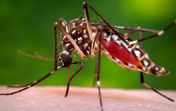
EEID scientists will study the effects of temperature on vector-borne disease transmission.
Credit and Larger Version

August 26, 2015

Emerging pandemic disease outbreaks such as Ebola increasingly threaten global public health and world economies, scientists say. We can expect five such new diseases to emerge each year--and spread. The tropical disease dengue fever, for example, has made its way to Florida and Texas, seemingly to stay.

Is our interaction with the environment somehow responsible for the increase in incidence of these diseases? A joint program of the National Science Foundation (NSF), National Institutes of Health (NIH) and the U.S. Department of Agriculture (USDA) is seeking answers.

The Ecology and Evolution of Infectious Diseases (EEID) program supports efforts to understand the underlying ecological and biological mechanisms behind human-induced environmental changes and the emergence and transmission of infectious diseases.

A complex process

The EEID program is also co-funded by the U.K.'s Biotechnology and Biological Sciences Research Council (BBSRC).

This year, the program has awarded eight new grants totaling $18 million.

Disease transmission is a complex process that involves disease organisms, disease vectors, disease hosts and the predators that consume those hosts. It links relatively pristine areas with human habitations and human-dominated areas.

Projects supported through the EEID program allow scientists to study how large-scale environmental events--such as habitat destruction, invasions of non-native species and pollution--alter the risks of emergence of viral, parasitic and bacterial diseases in humans and other animals.

Researchers supported through the EEID program are advancing basic theory related to infectious diseases, and applying that knowledge to improve our understanding of how pathogens spread through populations at a time of increasing global change.

EEID research benefits

The benefits of research on the ecology of infectious diseases include development of theories of how diseases are transmitted; improved understanding of unintended health effects of development projects; increased capacity to forecast disease outbreaks; and knowledge of how infectious diseases emerge and reemerge.

"As demonstrated by the Ebola crisis, infectious diseases are an ongoing threat," says Sam Scheiner of NSF's Directorate for Biological Sciences and EEID program officer at NSF. "The fundamental research from these projects will help prepare us for the next outbreak, wherever it might come from."

Adds Christine Jessup of NIH's Fogarty International Center, "Infectious diseases are an ongoing global health challenge, often with devastating consequences. Environmental change, population mobility, and complex socio-ecological systems underlie many infectious disease threats. Our ability to prevent and control emerging and re-emerging diseases hinges on enhanced understanding of these diseases in their ecological and evolutionary contexts."

New awards to address current and future threats

This year's EEID awardees will conduct research on such topics as: group living as a possible explanation for infectious disease vulnerability in social species; vector behavior in transmission ecology; effects of agricultural expansion and intensification on infections; long-distance dispersal and disease outbreaks; and effects of temperature on vector-borne disease transmission.

"As we learn more about the ecology of pathogens that cause infectious diseases, we see clear links among public health, animal health, plant health, and the environment, with agriculture playing a significant role," says Sonny Ramaswamy, USDA's National Institute of Food and Agriculture director. "Through our partnership with the Ecology and Evolution of Infectious Diseases program, we are able to support agriculturally-relevant research on topics of global concern, and help ensure the safety and security of our food supply."

Adds Melanie Welham, BBSRC's science director, "Global uncertainties can present new challenges, and scientific research helps us to prepare for our future. The health of our livestock, plants and crops is dependent on improved knowledge of infectious diseases. This new funding will help us respond more rapidly and effectively to emerging threats, and to safeguard health and food security."

2015 NSF-NIH-USDA-BBSRC Ecology and Evolution of Infectious Diseases Awards

Kathleen Alexander, Virginia Polytechnic Institute and State University: Can group living and the influence of Allee Effects explain infectious disease vulnerability in social species? Emergence of M. mungi in the cooperative breeding banded mongoose

Jason Blackburn, University of Florida: Spatio-temporally explicit estimation of R0 for pathogens with environmentally-mediated transmission

Sonia Hernandez, University of Georgia: Consequences of Anthropogenic Resources for the Cross-Scale Dynamics of an Enteric Pathogen in an Avian Host

Leah Johnson, University of South Florida: US-UK Collab: RCN: Vector Behavior in Transmission Ecology (VectorBiTE)

Cristina Lanzas, North Carolina State University-Raleigh: Exposure heterogeneity and environmental transmission dynamics of Escherichia coli

Erin Mordecai, Stanford University: Effects of temperature on vector-borne disease transmission: integrating theory with empirical data

Christopher Mundt, Oregon State University: Long-Distance Dispersal and Disease Outbreaks: Effects of Initial Prevalence, Basic Reproduction Number, and Control Tactics

Jason Rohr, University of South Florida: Effects of Agricultural Expansion and Intensification on Infections

-NSF-

Media Contacts
Cheryl Dybas, NSF, (703) 292-7734,
cdybas@nsf.gov

Related Websites
NSF Special Report: Ecology and Evolution of Infectious Diseases:http://www.nsf.gov/news/special_reports/ecoinf/
NSF Discovery Article Series: Ecology and Evolution of Infectious Diseases:http://nsf.gov/discoveries/disc_summ.jsp?cntn_id=134947
NSF News (2014 EEID Awards): Racing ahead of disease outbreaks: $12 million in new research grants:http://nsf.gov/news/news_summ.jsp?cntn_id=132570
NSF News (2013 EEID Awards): Outbreak: Ecology and Evolution of Infectious Disease grants support research on disease transmission:http://www.nsf.gov/news/news_summ.jsp?cntn_id=129280
NSF News (2012 EEID Awards): Controlling the Spread of Diseases Among Humans, Other Animals and the Environment:http://www.nsf.gov/news/news_summ.jsp?cntn_id=125496

Lemon juice disinfects against human norovirus


Noroviruses are the predominant cause of gastroenteritis outbreaks in community settings such as hospitals, cruise ships, and schools. The virus is extremely contagious and is mostly transmitted via "fecal-oral-route," i.e., through contaminated hands or contaminated food. Symptoms include violent and sudden onset of diarrhea, vomiting, and nausea.

"It is therefore important to provide a safe and harmless disinfectant against human norovirus," explains Grant Hansman, head of CHS junior research group at the German Cancer Research Center noroviruses and the University of Heidelberg. The group is funded by the CHS Foundation. Some older observations found that fruit extracts, such as orange or pomegranate juice, could reduce the infectivity of surrogate noroviruses.

This current study was a continuation of an earlier project conducted at the National Institutes of Health in the United States, where they discovered that citrate from a commercial company could bind to the norovirus capsid protein. "However, it was only by chance that this discovery was made, like a lot of scientific research, but it turns out to be very interesting." Dr Hansman explains.

Human noroviruses does not grow in cell culture, so they tested the effects of increasing concentrations of citric buffer on the so called norovirus virus-like particles, which have the same surface characteristics as real viruses. Dr Hansman and his staff show that the virus particles change their shape after citrate binding. X-ray crystal structures revealed that the citrate -- from lemon juice or citrate disinfectants -- precisely interacts at the binding pocket on the particle that is involved in attaching to host ligands -- the so-called histo-blood group antigens.

These new results may explain why citrate reduces the infectivity of noroviruses. "Maybe a few drops of lemon juice on contaminated food or surfaces may prevent the transmission of these viruses," speculates Hansman. With his staff, he now plans to investigate if citric acid could reduce symptoms in those already infected with noroviruses.


Story Source:

The above post is reprinted from materials provided by German Cancer Research Center (Deutsches Krebsforschungszentrum, DKFZ). Note: Materials may be edited for content and length.


Journal Reference:

  1. Anna D. Koromyslova, Peter A. White, Grant S. Hansman. Treatment of norovirus particles with citrate. Virology, 2015; 485: 199 DOI:10.1016/j.virol.2015.07.009


Top 30 Most Beautiful and Eye-capturing Earth Photographs of 2015


Everyone loves nature and natural photographs as well. People travel everywhere and capture the most beautiful and perfect photographs for their collection or projects. Some of our friends click some amazing photographs of natural beauty which attracts deeply and we want to visit that place.

Top 30 Most Beautiful and Eye-capturing Earth Photographs of 2015

These photographs are taken by different people at different places, but the one thing which is common on these is the originality and beauty of nature.
Today we collect some stunning and beautiful photographs of Natural Beauty of Earth, you must enjoy them.

Nordaustlandet, Svalbard, Norway

Top 30 Most Beautiful and Eye-capturing Earth Photographs of 2015

Mt. McKinley completely visible with a reflection!

Top 30 Most Beautiful and Eye-capturing Earth Photographs of 2015

Antelope Canyon

Top 30 Most Beautiful and Eye-capturing Earth Photographs of 2015

Ice on Lake Baikal

Top 30 Most Beautiful and Eye-capturing Earth Photographs of 2015

Twelve Apostles, Australia

Top 30 Most Beautiful and Eye-capturing Earth Photographs of 2015

Grand Canyon National Park, Arizona

Top 30 Most Beautiful and Eye-capturing Earth Photographs of 2015

View of Sedona from End of the World, Flagstaff, AZ

Top 30 Most Beautiful and Eye-capturing Earth Photographs of 2015

Sun sets over the Fitz Roy, Patagonia, Chile

Top 30 Most Beautiful and Eye-capturing Earth Photographs of 2015

Mirror Lake, Yosemite National Park.

Top 30 Most Beautiful and Eye-capturing Earth Photographs of 2015

Half Dome, Yosemite National Park, California

Top 30 Most Beautiful and Eye-capturing Earth Photographs of 2015

Belgium by Chung Hu

Top 30 Most Beautiful and Eye-capturing Earth Photographs of 2015

Piscinas Naturales De Garachico in Tenerife, Spain

Top 30 Most Beautiful and Eye-capturing Earth Photographs of 2015

Antelope Canyon

Top 30 Most Beautiful and Eye-capturing Earth Photographs of 2015

Taos Gorge in New Mexico

Top 30 Most Beautiful and Eye-capturing Earth Photographs of 2015

Delta Lake, WY

Top 30 Most Beautiful and Eye-capturing Earth Photographs of 2015

Summer in England

Top 30 Most Beautiful and Eye-capturing Earth Photographs of 2015

East Greenland Coast

Top 30 Most Beautiful and Eye-capturing Earth Photographs of 2015

Dolomites.

Top 30 Most Beautiful and Eye-capturing Earth Photographs of 2015

Owl Creek Pass, Colorado

Top 30 Most Beautiful and Eye-capturing Earth Photographs of 2015

A forbidden hill in Ireland

Top 30 Most Beautiful and Eye-capturing Earth Photographs of 2015

Mt. Fuji, Japan

Top 30 Most Beautiful and Eye-capturing Earth Photographs of 2015

Corinth, Greece.

Top 30 Most Beautiful and Eye-capturing Earth Photographs of 2015

The Coeur d'Alene River

Top 30 Most Beautiful and Eye-capturing Earth Photographs of 2015

Grand Canyon, North Rim

Top 30 Most Beautiful and Eye-capturing Earth Photographs of 2015

El Capitan just after sunrise Guadalupe Mountains

Top 30 Most Beautiful and Eye-capturing Earth Photographs of 2015

South Rim of the Grand Canyon, Arizona

Top 30 Most Beautiful and Eye-capturing Earth Photographs of 2015

Morning on the Lost Coast

Top 30 Most Beautiful and Eye-capturing Earth Photographs of 2015

A perfect morning in Big Sur.

Top 30 Most Beautiful and Eye-capturing Earth Photographs of 2015

Lake Limedes, under the Lagazuoi in the Dolomites

Top 30 Most Beautiful and Eye-capturing Earth Photographs of 2015


http://www.topntoday.co/2015/07/top-30-most-beautiful-and-eye-capturing.html