domingo, 18 de maio de 2014

Magnets and kids: A dangerous duo


Magnet ingestions by children have received increasing attention over the past 10 years. With the growing availability of new and stronger neodymium-iron-boron magnets being sold as "toys," there has been an increase of cases of ingestion, resulting in serious injury and, in some cases, death. In a new study scheduled for publication in The Journal of Pediatrics, researchers studied the trends of magnetic ingestions at The Hospital for Sick Children (SickKids), Canada's largest children's hospital.

Matt Strickland, MD, and colleagues reviewed all cases of foreign body ingestion seen in the emergency department from April 1, 2002 through December 31, 2012. Inclusion criteria included being less than 18 years of age, with suspected or confirmed magnetic ingestion. According to Dr. Strickland, "We chose to limit our scope to the alimentary tract because the majority of serious harm from magnets arises from perforations and fistulae of the stomach, small bowel, and colon." To reflect the introduction of small, spherical magnet sets in 2009, the study was divided into two time periods, visits during 2002-2009 and those during 2010-2012.

Of 2,722 patient visits for foreign body ingestions, 94 children met the inclusion criteria. Of those, 30 children had confirmed ingestion of multiple magnets. Overall magnet ingestions tripled from 2002-2009 to 2010-2012; the incidence of injuries involving multiple magnets increased almost 10-fold between the two time periods. Six cases required surgery for sepsis or potential for imminent bowel perforation, all of which occurred in 2010-2012. The average size of the magnets also decreased approximately 70% between 2002-2009 and 2010-2012.

This study shows a significant increase in the rate of multiple magnet-related injuries between 2002 and 2012. "More concerning, however," notes Dr. Strickland, "is the increased number of high-risk injuries featuring multiple, smaller magnets." Despite new magnet-specific toy standards, labeling requirements, product recalls, and safety advisories issued in the past 10 years, continuing efforts should focus on educating parents and children on the dangers inherent in magnetic "toys."


Story Source:

The above story is based on materials provided by Elsevier. Note: Materials may be edited for content and length.

Cameras and displays: Organic photodiodes for sensor applications


Organic photodiodes are extremely lightweight and also inexpensive to produce.

Powerful, inexpensive and even flexible when they need to be, organic photodiodes are a promising alternative to silicon-based photodetectors. They are used to improve light sensitivity in cameras and to check displays for homogeneous color composition. Fraunhofer scientists are developing just this kind of component to fit customer-specific requirements and will be presenting a color sensor demonstrator at this year's SENSOR + TEST trade fair in Nuremberg from June 3 to 5.

We have Mother Nature and her ingenious brand of technology to thank for our ability to see -- our eyes capture light from our environment and our retinas transform it into an electronic signal that is passed on to the brain as information. Optical components -- like photodetectors -- work according to the same principle. They can be found in digital cameras and are also used in automation technology, bioanalytics and medical imaging diagnostics. Optical components are typically made from inorganic materials such as silicon. But scientists at the Fraunhofer Research Institution for Organics, Materials and Electronic Devices COMEDD in Dresden are now also developing organic photodiodes (OPD) that rely on organic materials such as dyes or pigments. "These kinds of OPD offer a range of advantages compared with inorganic components -- they're extremely lightweight, cheap to produce and can be used for flexible applications," explains COMEDD head of department Dr. Olaf R. Hild.

The choice of which material to use is determined primarily by the wavelength spectrum customers select for their applications. Organic materials are each sensitive only to a particular wavelength range -- for instance, they may react only to green light. So by choosing the right material, scientists can control and tailor the spectral sensitivity of their optical sensors. The available materials already cover a broad wavelength spectrum. For special applications, for instance in the UV or near-infrared range, the Dresden-based scientists are also developing compact micro-sensors that combine organic semiconductors with silicon technology.

Increased light sensitivity using photodiodes

Uses vary from tiny sensor elements for cameras or for bioanalytics to large-scale, quality control applications. In lab-on-chip applications, for instance, OPDs can detect certain DNA sequences that have been tagged with fluorescent markers. Hild explains how photodiodes help to increase a high-end camera's light sensitivity: "Integrating our organic photodiodes increases the light sensitivity of today's CCD chips by providing a larger usable surface." OPDs can also be used to check the homogeneity of the color composition or the brightness distribution of luminescent surfaces such as displays.

Unlike their silicon-based counterparts, OPDs also allow for flexible components. Here photodiodes are integrated into polymer films that can be applied to concave or curved surfaces. This could be used to develop quality control systems specially shaped to the product to be placed inside them so that whole car doors could be examined for scratches or any quality inconsistencies in the paintwork. And it is particularly in such large-scale applications that OPDs offer a cheaper alternative to traditional technologies: whereas it is very difficult and expensive to cover large surfaces with silicon, OPDs can be applied to comparatively inexpensive materials using simple coating techniques. This means that the scientists can utilize established manufacturing techniques such as those used to manufacture organic photovoltaics, for example. At this year's SERSOR + TEST the scientists will be presenting a color sensor featuring four organic photodiodes, each one with its own spectral sensitivity.


Story Source:

The above story is based on materials provided by Fraunhofer-Gesellschaft. Note: Materials may be edited for content and length.
Silently among us: Scientists worry about milder cases of MERS


(Reuters) - Scientists leading the fight against Middle East Respiratory Syndrome say the next critical front will be understanding how the virus behaves in people with milder infections, who may be spreading the illness without being aware they have it.

Establishing that may be critical to stopping the spread of MERS, which emerged in the Middle East in 2012 and has so far infected more than 500 patients in Saudi Arabia alone. It kills about 30 percent of those who are infected.

It is becoming increasingly clear that people can be infected with MERS without developing severe respiratory disease, said Dr David Swerdlow, who heads the MERS response team at the U.S. Centers for Disease Control and Prevention.

"You don't have to be in the intensive care unit with pneumonia to have a case of MERS," Swerdlow told Reuters. "We assume they are less infectious (to others), but we don't know."

The CDC has a team in Saudi Arabia studying whether such mild cases are still capable of spreading the virus. Swerdlow is overseeing their work from Atlanta.

They plan to test the family members of people with mild MERS, even if these relatives don't have any symptoms, to help determine whether the virus can spread within a household.

Cases of the disease, which causes coughing, fever and sometimes fatal pneumonia, have nearly tripled in the past month and a half, and the virus is moving out of the Arabian peninsula as infected individuals travel from the region.

Since late April, the first two cases of MERS have been reported on U.S. soil. Dutch officials reported their first two cases this week. Infections have also turned up in Britain, Greece, France, Italy, Malaysia and elsewhere.

Since MERS is an entirely new virus, there are no drugs to treat it and no vaccines capable of preventing its spread. It is a close cousin of the virus that caused Severe Acute Respiratory Syndrome or SARS, which killed around 800 people worldwide after it first appeared in China in 2002.

Because MERS patients can have "mild and unusual symptoms," the World Health Organization advises healthcare workers to apply standard infection control precautions for all patients, regardless of their diagnosis, at all times.

"Asymptomatic carriers of diseases can represent a major route for a pathogen to spread," said Dr Amesh Adalja of the University of Pittsburgh Medical Center.

"Just think of Typhoid Mary," he said, referring to the asymptomatic cook who spread typhoid fever to dozens of people in the early 20th century.

NOT EVEN A COUGH

Milder symptoms played a role in the second U.S. case of MERS, a man who started having body aches on a journey from Jeddah on Saudi Arabia's Red Sea coast to the United States.

It took the patient more than a week before he sought help in an emergency department in Orlando, Florida. Once he arrived, he waited nearly 12 hours in the ER before staff recognized a MERS link and placed him in an isolation room. The patient did not have signs of a respiratory infection, not even a cough.[ID:nL1N0O002W]

Dr Kevin Sherin, director of the Florida Department of Health for Orange County, believes that made it less likely that he could spread the infection. Hospital workers have tested negative, but the health department and the CDC are still checking on hundreds of people who might have been in contact with the patient.

A CDC study published earlier this week looked at some of the first cases of MERS that occurred in Jordan in 2012.

Initially, only two people in that outbreak were thought to have MERS. When CDC disease detectives used more sensitive tests that looked for MERS antibodies among hospital workers, they found another seven people had contracted MERS and survived it.

That suggests there may be people with mild cases "that can serve as a way for the virus to spread to other individuals, which makes it a lot harder to control," Adalja said.

Scientists are especially concerned because a lot of recent cases of MERS are among people who did not have contact with animals such as camels or bats that are believed to be reservoirs for the virus.

"If they don't have animal contact, where do they pick it up? Potentially, asymptomatic cases," said Dr Michael Osterholm, an infectious disease expert from the University of Minnesota.

Herpes-loaded stem cells used to kill brain tumors


Harvard Stem Cell Institute (HSCI) scientists at Massachusetts General Hospital have a potential solution for how to more effectively kill tumor cells using cancer-killing viruses. The investigators report that trapping virus-loaded stem cells in a gel and applying them to tumors significantly improved survival in mice with glioblastoma multiforme, the most common brain tumor in human adults and also the most difficult to treat.

The work, led by Khalid Shah, MS, PhD, an HSCI Principal Faculty member, is published in the Journal of the National Cancer Institute. Shah heads the Molecular Neurotherapy and Imaging Laboratory at Massachusetts General Hospital.

Cancer-killing or oncolytic viruses have been used in numerous phase 1 and 2 clinical trials for brain tumors but with limited success. In preclinical studies, oncolytic herpes simplex viruses seemed especially promising, as they naturally infect dividing brain cells. However, the therapy hasn't translated as well for human patients. The problem previous researchers couldn't overcome was how to keep the herpes viruses at the tumor site long enough to work.

Shah and his team turned to mesenchymal stem cells (MSCs) -- a type of stem cell that gives rise to bone marrow tissue -- which have been very attractive drug delivery vehicles because they trigger a minimal immune response and can be utilized to carry oncolytic viruses. Shah and his team loaded the herpes virus into human MSCs and injected the cells into glioblastoma tumors developed in mice. Using multiple imaging markers, it was possible to watch the virus as it passed from the stem cells to the first layer of brain tumor cells and subsequently into all of the tumor cells.

"So, how do you translate this into the clinic?" asked Shah, who also is an Associate Professor at Harvard Medical School.

"We know that 70-75 percent of glioblastoma patients undergo surgery for tumor debulking, and we have previously shown that MSCs encapsulated in biocompatible gels can be used as therapeutic agents in a mouse model that mimics this debulking," he continued. "So, we loaded MSCs with oncolytic herpes virus and encapsulated these cells in biocompatible gels and applied the gels directly onto the adjacent tissue after debulking. We then compared the efficacy of virus-loaded, encapsulated MSCs versus direct injection of the virus into the cavity of the debulked tumors."

Using imaging proteins to watch in real time how the virus combated the cancer, Shah's team noticed that the gel kept the stem cells alive longer, which allowed the virus to replicate and kill any residual cancer cells that were not cut out during the debulking surgery. This translated into a higher survival rate for mice that received the gel-encapsulated stem cells.

"They survived because the virus doesn't get washed out by the cerebrospinal fluid that fills the cavity," Shah said. "Previous studies that have injected the virus directly into the resection cavity did not follow the fate of the virus in the cavity. However, our imaging and side-by-side comparison studies showed that the naked virus rarely infects the residual tumor cells. This could give us insight into why the results from clinical trials with oncolytic viruses alone were modest."

The study also addressed another weakness of cancer-killing viruses, which is that not all brain tumors are susceptible to the therapy. The researchers' solution was to engineer oncolytic herpes viruses to express an additional tumor-killing agent, called TRAIL. Again, using mouse models of glioblastoma -- this time created from brain tumor cells that were resistant to the herpes virus -- the therapy led to increased animal survival.

"Our approach can overcome problems associated with current clinical procedures," Shah said. "The work will have direct implications for designing clinical trials using oncolytic viruses, not only for brain tumors, but for other solid tumors."

Further preclinical work will be needed to use the herpes-loaded stem cells for breast, lung and skin cancer tumors that metastasize to the brain. Shah predicts the approach will enter clinical trials within the next two to three years.


Story Source:

The above story is based on materials provided by Harvard University. Note: Materials may be edited for content and length.


Journal Reference:

  1. Matthias Duebgen, Jordi Martinez-Quintanilla, Kaoru Tamura, Shawn Hingtgen, Navid Redjal, Hiroaki Wakimoto And Khalid Shah. Stem Cells Loaded With Multimechanistic Oncolytic Herpes Simplex Virus Variants for Brain Tumor Therapy. Journal of the National Cancer Institute, May 2014 DOI: 10.1093/jnci/dju090

Loneliness Hurts the Heart


How this chronic form of stress can lead to heart disease

lonely-hearts

(GETTYIMAGES)

Being alone can break your heart—literally.

People who lack a strong network of friends and family are at greater risk of developing—and dying from—heart disease, research shows. According to some studies, the risk of solitude is comparable to that posed by high cholesterol, high blood pressure, and even smoking.

Experts havent pinpointed exactly how social networks protect against heart disease, but there are a number of probable explanations. People who are socially isolated are more likely to drink, smoke, and get less exercise. And once someone has heart disease, friends and family often provide key support, such as picking up prescriptions, encouraging exercise, cooking healthy meals, and helping with household chores.
While that everyday help is important, its not the whole story. In recent years, researchers have begun to unravel the cardiovascular effects of social isolation, and theyve discovered that feeling alone may hurt the heart even more than actually being alone.
“We started looking at social isolation about 20 years ago, and we found fairly quickly that objective social isolation in everyday life isnt as important as perceived social isolation,” says John Cacioppo, PhD, a professor of psychology at the University of Chicago. “And theres a term for perceived social isolation: Its loneliness.”
What we call loneliness—the feeling that you have no one to turn to, that no one understands you—is a form of stress. And if it becomes chronic, it can wreak havoc on your blood vessels and heart.
What is loneliness?
Though the concepts are sometimes used interchangeably, loneliness is distinct from social isolation (also known as low social support). Theres some overlap between the two, but not as much as one might think.
Social support is typically measured using a handful of characteristics such as marital status, number of friends, and participation in group activities (such as churchgoing). Low scores on these measures dont necessarily correspond to loneliness, however. Some people need more “me” time than others, after all, and some people are content with just one or two close friends.
To factor in these individual preferences, researchers define loneliness as the gap between a persons desired and actual social relationships—a subjective measure thats most easily gauged with questionnaires. To put it another way, social isolation mainly describes the extent of a persons social network, while loneliness emphasizes quality, rather than quantity, and describes the satisfaction and comfort a person derives from their interpersonal relationships. Its the difference between the amount of food on your plate and how good it tastes.
Fifteen years ago, after undergoing surgery to replace a heart valve, Dale Briggs, 63, of Clovis, Calif., felt anxious and isolated, not like himself at all. Briggs rated high on the social support scale—hes married, hes a weekly churchgoer—but he felt that he wasnt connecting with people anymore.
“During that period I could have had 15 people in my house all the time, but I would have traded them all for someone who had been through what Id been through and who I could talk to about it,” Briggs recalls. “I felt isolated in my mind, like I couldnt relate to anybody.”

High dose of radiation on heart attack patients.


Heart Tests|Study: Heart Attack Patients Receive High Dose of Radiation - Heart Disease - Health.com

Belly fat.


Why You're Not Losing Belly Fat - Health.com

Neuroglial Cells


Neuroglial cells—usually referred to simply as glial cells or glia—are quite different from nerve cells. The major distinction is that glia do not participate directly in synaptic interactions and electrical signaling, although their supportive functions help define synaptic contacts and maintain the signaling abilities of neurons. Glia are more numerous than nerve cells in the brain, outnumbering them by a ratio of perhaps 3 to 1. Although glial cells also have complex processes extending from their cell bodies, they are generally smaller than neurons, and they lack axons and dendrites. The term glia (from the Greek word meaning “glue”) reflects the nineteenth-century presumption that these cells held the nervous system together in some way. The word has survived, despite the lack of any evidence that binding nerve cells together is among the many functions of glial cells. Glial roles that are well-established include maintaining the ionic milieu of nerve cells, modulating the rate of nerve signal propagation, modulating synaptic action by controlling the uptake of neurotransmitters, providing a scaffold for some aspects of neural development, and aiding in (or preventing, in some instances) recovery from neural injury.

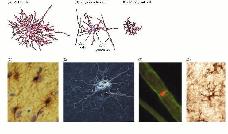
Neuroglial cells. Tracings of an astrocyte (A), an oligodendrocyte (B), and a microglial cell (C) visualized by impregnation with silver salts. The images are at approximately the same scale. (D) Astrocytes in the brain labeled with an antibody against

Figure 1.4. Neuroglial cells.

There are three types of glial cells in the mature central nervous system: astrocytes, oligodendrocytes, and microglial cells .Astrocytes, which are restricted to the brain and spinal cord, have elaborate local processes that give these cells a starlike appearance (hence the prefix “astro”). The major function of astrocytes is to maintain, in a variety of ways, an appropriate chemical environment for neuronal signaling. Oligodendrocytes, which are also restricted to the central nervous system, lay down a laminated, lipid-rich wrapping called myelin around some, but not all, axons. Myelin has important effects on the speed of action potential conduction . In the peripheral nervous system, the cells that elaborate myelin are called Schwann cells. As the name implies, microglial cells are smaller cells derived from hematopoietic stem cells (although some may be derived directly from neural stem cells). They share many properties with tissue macrophages, and are primarily scavenger cells that remove cellular debris from sites of injury or normal cell turnover. Indeed, some neurobiologists prefer to categorize microglia as a type of macrophage. Following brain damage, the number of microglia at the site of injury increases dramatically. Some of these cells proliferate from microglia resident in the brain, while others come from macrophages that migrate to the injured area from the circulation.