quarta-feira, 7 de maio de 2014

Why Some Doctors Like Google Glass So Much


The way some emergency doctors are using Glass highlights the promise, and the limitations, of wearable technology.

Why It Matters

Access to patient records and other medical data can help doctors save lives.

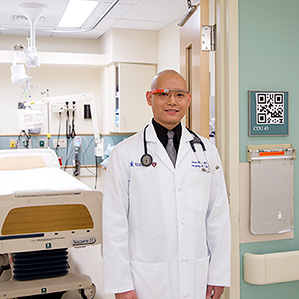
Digital doc: Emergency physician and clinical informaticist Steve Horng wears the Google Glass device used in Beth Israel Deaconess Medical Center’s emergency department.

Kermit the Frog showed up in the emergency room at Beth Israel Deaconess Medical Center in Boston recently, complaining of chest pain. A quick tilt of my head showed me Kermit’s records—his EKG results, the radiology tests ordered for him, and his medical history.

Don’t worry, Kermit’s not really sick. The frog’s emergency room visit was just meant to illustrate how Google’s face-mounted computer, Glass, can let physicians quickly get up to speed on a patient’s situation without having to turn repeatedly to a computer.

Physicians in the hospital’s emergency department are in the midst of a pilot project using heavily modified versions of Glass to look up patient records. Emergency physician Steve Horng is spearheading the project, which offers Glass to all physicians in the department. “Emergency medicine is a very information-intensive specialty where even small nuggets of information available immediately really matter,” he says. “Having information one minute earlier can actually be quite life-saving.”

The experiment is one of many tests to see if face-worn computers can be beneficial for workers who need fast access to small amounts of information without taking their hands or even their full gaze away from other tasks. These efforts hint at the potential benefits, as well as the remaining challenges, of wearable computing in general.

Google isn’t the only company offering devices that meet these needs. For example, Epson sells a pair of goggles that help nurses see veins through a patient’s skin, and Vuzix produces head-mounted displays for the defense industry that can identify friendly forces and more (see “Hands-On with the Vuzix M100, a Google Glass Competitor”).

The emergency room team at Beth Israel has four pairs of Glass, which can be picked up at the beginning of a shift. After a nurse has checked a patient in, a doctor uses Glass to scan a QR code on the outside of the patient’s room. That simply tells the device which room number the doctor is entering; the custom app then looks up patient records on the hospital Wi-Fi network and displays the records to the doctor on a small prism in front of one eye.

The amount of information available through the app is limited, and it’s not possible to do complicated searches or any data entry with gesture and voice commands. But for emergency medicine, the high-level facts are useful, says Horng.

San Francisco-based Wearable Intelligence developed a custom application for Glass that blocks all the device’s social-media features and locks it to the hospital’s Wi-Fi network. Wearable Intelligence has developed other custom applications for industrial uses of Glass, such as one that provides real-time contextual information for oil and gas workers.

While Glass has its skeptics (see “Glass, Darkly” and “Google Glass Still Needs a Killer App”), there is some evidence the technology can be helpful in industries like these. At a recent event hosted by Google’s Cambridge branch, doctors from across the country came to show off how they’d thought of harnessing Glass for medicine. One presenter, Rafael Grossman, a surgeon based in Bangor, Maine, was the first person to use Glass during live surgery. He thinks the technique could help doctors teach new surgeons.

But for the pilot at Beth Israel, video is off the table, at least for now. “We wanted to stay away from anything that could potentially be misconstrued as leaking patient information, so until we had a case study and a good foundation, we purposely stayed away from enabling the video feed,” says Horng.

Medical gaze: A screenshot of a patient’s health records as seen through Beth Israel Deaconess Medical Center’s modified Google Glass.

Clearly, Glass still has limitations. When I visited Horng at Beth Israel, a collaborator from Wearable Intelligence was testing out a new external battery pack for the device. Without the extra boost, Glass can last about two hours running the processing-intensive medical app, says Horng. That’s inadequate given the eight-hour shifts in the emergency department.

Like people in the wider population, some doctors doubt Glass’s usefulness. Emergency physicians in general are very technology savvy, Horng says, but they vary in their enthusiasm for the pilot project. “You’ve got the really early adopters that will try anything and just like new technology, and then you’ve got the other side that just refuses to get away from their clipboard, and so they are never going to use it,” he says.

But plenty of doctors seem excited, and they do not see Glass as a barrier to patient-doctor interaction. In fact, says Karandeep Singh, a nephrologist at Brigham and Women’s Hospital in Boston, they believe Glass could help improve it. “The advent of electronic health records has significantly changed that [doctor-patient] relationship,” he told the audience at Google’s Cambridge event. A lot of times, doctors equate looking at electronic records with seeing patients, said Singh: “Some of the art of medicine has been lost.” Glass offers a way to look up important patient data without breaking contact at the bedside, he said.

In the next version of the Glass app used at Beth Israel, the team plans to add voice commands, probably using a non-Google voice recognition engine. “Right now Google’s voice recognition isn’t as resilient as we would like,” says Horng.

The limitations of the Glass interface also represent a new area for potential innovation. “The use of mobile devices and all the opportunities that that opens up in clinical care create new needs on the informatics and algorithms side,” says David Sontag, a computer scientist at NYU, who is collaborating with Horng. “Only a small amount of information would be visible to the clinician. There simply aren’t that many pixels to display on, and time is of the essence.”

What will make Glass and other wearable devices more useful will be algorithms that can get the right data to doctors at the right time, says Sontag. “They are simply even more important now in the context of mobile devices.”

FVD Speed Dial - Mozilla Firefox 2014-03-22 22.47.08

Nanotube-Infused Clothing May Protect Against Chemical Weapons


From NIST Tech Beat: May 6, 2014


Nerve agents are among the world's most feared chemical weapons, but scientists at the National Institute of Standards and Technology (NIST) have demonstrated a way to engineer carbon nanotubes to dismantle the molecules of a major class of these chemicals.* In principle, they say, the nanotubes could be woven into clothing that destroys the nerve agents on contact before they reach the skin.

The team's experiments show that nanotubes—special molecules that resemble cylinders formed of chicken wire—can be combined with a copper-based catalyst able to break apart a key chemical bond in the class of nerve agents that includes Sarin. A small amount of catalyst can break this bond in a large number of molecules, potentially rendering a nerve agent far less harmful. Because nanotubes further enhance the breakdown capability of the catalyst and can be woven into fabric easily, the NIST team members say the findings could help protect military personnel involved in cleanup operations.

Sarin—used in a 1995 Tokyo subway attack—is one of several deadly nerve agents of a group called organophosphates. Many are classified as weapons of mass destruction. While organophosphates are harmful if inhaled, they also are dangerous if absorbed through the skin, and can be even be re-released from clothing if not thoroughly decontaminated.

To protect themselves during research, the team did not work with actual nerve agents, but instead used a "mimic molecule" that contains a chemical bond identical to the one found in organophosphates. Breaking this bond splits the molecule into pieces that are far less dangerous.

The team developed a way to attach the catalyst molecule to the nanotubes and then tested the effectiveness of the tube-catalyst complex to break the bonds. To perform the test, the complex was deposited onto a small sheet of paper and put into a solution containing the mimic molecule. For comparison, the catalyst without nanotubes was tested simultaneously in a different solution. Then it was a simple matter of stirring and watching chemistry in action.

"The solution was initially transparent, almost like water," says the team's John Heddleston, "but as soon as we added the paper, the solution started to turn yellow as the breakdown product accumulated. Measuring this color change over time told us the amount and rate of catalysis. We began to see a noticeable difference within an hour, and the longer we left it, the more yellow it became." The catalyst-nanotube complex far outperformed the catalyst alone.

Principal investigator Angela Hight Walker says that several questions will need to be addressed before catalytic nanotubes start showing up in clothing, such as whether it is better to add the catalyst to the nanotubes before or after they are woven into the fabric.

"We'd also like to find ways to make the catalytic reaction go faster, which is always better," Hight Walker says. "But our research group has been focusing on the fundamental science of nanoparticles for years, so we are in a good position to answer these questions."

*M.M. Bailey, J.M. Heddleston, J. Davis, J.L. Staymates and A.R. Hight Walker. Functionalized, carbon nanotube material for the catalytic degradation of organophosphate nerve agents. Nano Research, DOI:10.1007/s12274-014-0405-3, Nano Research 2014, 7(3): 390–398.

Impulsivity is risk factor for food addiction


May 6, 2014

Boston University Medical Center

Have you ever said to yourself that you would only have a handful of potato chips from the bag then, minutes later, realized you ate the whole thing? A recent study shows that this type of impulsive behavior might not be easily controlled -- and could be a risk factor in the development of food addiction and eating disorders as a result of cellular activities in the part of the brain involved with reward.


The research was led by Boston University School of Medicine (BUSM) and conducted in collaboration with the University of Cambridge in the United Kingdom. It also points out the common mechanisms involved between drug and food addiction. (Pictured L-R: Dr. Valentina Sabino, Cassie Moore, Dr. Clara Velazquez, Toni Ferragud and Dr. Pietro Cottone)

Have you ever said to yourself that you would only have a handful of potato chips from the bag then, minutes later, realized you ate the whole thing? A recent study shows that this type of impulsive behavior might not be easily controlled -- and could be a risk factor in the development of food addiction and eating disorders as a result of cellular activities in the part of the brain involved with reward.

Research has shown that people with eating disorders and obesity are known to be more impulsive than healthy people. For example, they may be more likely to blurt out something that they later regret saying or to start an activity without thinking through the consequences. However, it was unclear whether the impulsivity existed before the dysfunctional eating behavior or if developed as a result of it.

BUSM researchers attempted to answer this question by measuring the inability to withhold an impulsive response in experimental models that were exposed to a diet high in sugar daily for one hour. Models shown to be more impulsive rapidly developed binge eating, showing heightened cravings and the loss of control over the junk diet (measured as inability to properly evaluate the negative consequences associated with ingestion of the sugary diet). Conversely, models shown to be less impulsive demonstrated the ability to appropriately control impulsive behavior and did not show abnormal eating behavior when exposed to the sugary diet.

Interestingly, the impulsive models showed increased expression of a transcription factor called Delta-FosB in the nucleus accumbens, an area of the brain involved in reward evaluation and impulsive behavior, indicating a potential biological component to this behavior.

"While impulsivity might have aided ancestors to choose calorie-rich foods when food was scarce, our study results suggest that, in today's calorie-rich environment, impulsivity promotes pathological overeating," said Pietro Cottone, PhD, co-director of the Laboratory of Addictive Disorders and associate professor of pharmacology and psychiatry at BUSM.

"Our results add further evidence to the idea that there are similar mechanisms involved in both drug and food addiction behavior," said Clara Velazquez-Sanchez, PhD, postdoctoral fellow in the Laboratory of Addictive Disorder and first author of the study.


Story Source:

The above story is based on materials provided by Boston University Medical Center. Note: Materials may be edited for content and length.


Journal Reference:

  1. Clara Velázquez-Sánchez, Antonio Ferragud, Catherine F Moore, Barry J Everitt, Valentina Sabino, Pietro Cottone. High Trait Impulsivity Predicts Food Addiction-Like Behavior in the Rat. Neuropsychopharmacology, 2014; DOI: 10.1038/npp.2014.98

Ancient Egyptians likely used damp sand to help move pyramid-building blocks


Ancient Egyptians likely used damp sand to help move pyramid-building blocks

The miniature sledge-testing rig used in the study

The miniature sledge-testing rig used in the study

In Egypt's tomb of Djehutihotep, a wall painting depicts someone pouring water into the sand in front of one of the sledges that hauled the blocks used in the construction of the pyramids. According to new research, they had a good reason for doing so – by wetting the sand, as little as half as much pulling force would have been required to move those sledges.

The research was conducted by a team from the Foundation for Fundamental Research on Matter, and the University of Amsterdam. They built miniature versions of the laden sledges, which they pulled through trays filled with both dry and moistened sand.

When an optimum amount of water was added to dry sand, capillary action bonded the individual grains together, causing the material to become approximately twice as stiff as it had been. This stiffness kept the sand from piling up in front of the sledge, making it much easier to pull. Too much water, however, saturated the sand and caused it to lose that stiffness.

It's similar to the building of sand castles, in which damp sand holds together better than dry or sodden sand.

The painting from the tomb of Djehutihotep

The painting from the tomb of Djehutihotep

While there currently aren't many people attempting to build giant pyramids, the researchers believe that their findings could nonetheless have some practical applications. In particular, the knowledge could be used "to optimize the transport and processing of granular material" such as asphalt, concrete or coal.

A paper on the research was recently published in the journal Physical Review Letters.

Source: FOM Foundation

Linking vascular inflammation to obesity and atherosclerosis


New research reveals that IKKβ inhibitors reduce diet-induced obesity. These images show that fat mass is significantly decreased in mice treated with IKKβ inhibitors (right) compared with a control group (left).

A study in The Journal of Experimental Medicine shows that IκB kinase β (IKKβ) functions in smooth muscle cells to regulate vascular inflammatory responses and atherosclerosis development.

Inflammatory responses are the driving force of atherosclerosis, a process that involves the hardening and thickening of artery walls due to excess fatty deposits. IKKβ is a central coordinator of inflammatory responses that has been implicated in vascular diseases, but its role in atherosclerosis has been unclear.

Now, Changcheng Zhou and colleagues from the University of Kentucky show that deficiency of IKKβ in smooth muscle cells decreases vascular inflammation and atherosclerosis development in mice. Surprisingly, the lack of IKKβ also blocks the differentiation of fat cells and causes an accumulation of body fat precursor cells, thus protecting the animals from diet-induced obesity.

These novel findings suggest that the kinase acts as a regulator of fat cell differentiation. The use of IKKβ inhibitors may therefore provide an innovative treatment for atherosclerosis, obesity, and metabolic disorders.


Story Source:

The above story is based on materials provided by The Rockefeller University Press. Note: Materials may be edited for content and length.


Journal Reference:

  1. Y. Sui, S.-H. Park, J. Xu, S. Monette, R. N. Helsley, S.-S. Han, C. Zhou. IKK  links vascular inflammation to obesity and atherosclerosis. Journal of Experimental Medicine, 2014; 211 (5): 869 DOI: 10.1084/jem.20131281

College kids need to change unhealthy ways


May 6, 2014

Northwestern University

A new study has found that the majority of college students are engaging in unhealthy behaviors that could increase their risk of cancer later on. A shocking 95 percent of college students fail to eat the recommended amount of fruit and vegetables (five or more servings a day), and more than 60 percent report not getting enough physical activity (three or more days of vigorous exercise for at least 20 minutes or five or more days of moderate exercise for at least 30 minutes a week).


Parents, forget the comfort food! It's time to send your college students care packages of fruit, veggies and exercise gear instead.

A new study from Northwestern Medicine® and Northeastern Illinois University found that the majority of college students are engaging in unhealthy behaviors that could increase their risk of cancer later on. Racial minority students could be at an even greater risk, especially African Americans and Native Americans.

A shocking 95 percent of college students fail to eat the recommended amount of fruit and vegetables (five or more servings a day), and more than 60 percent report not getting enough physical activity (three or more days of vigorous exercise for at least 20 minutes or five or more days of moderate exercise for at least 30 minutes a week).

"Changing unhealthy behaviors in college students now could be a way to reduce the risk of cancer as well as other diseases later in life," said Brian Hitsman, principal investigator of the study.

Hitsman is an assistant professor in preventive medicine-behavioral medicine and psychiatry and behavioral sciences at Northwestern University Feinberg School of Medicine and a member of the Robert H. Lurie Comprehensive Cancer Center of Northwestern University.

Published online May 5 in the journal Preventive Medicine, the study is the first to evaluate cancer risk behaviors and conditions in college students and how they vary by race and ethnicity. Data for the study comes from the fall 2010 wave of the National College Health Assessment, a self-reported survey of a diverse group of more than 30,000 college students in the United States.

The majority of all college students surveyed reported low fruit and vegetable consumption and low physical activity. Other unhealthy behaviors or conditions -- alcohol binge drinking, tobacco use and obesity/being overweight -- appear to cluster differently among college students depending on their race, the scientists found.

For example, tobacco use and alcohol binge drinking seem to go hand in hand for all subgroups except black students. For black students, tobacco use co-occurred with being overweight/obese.

"Tobacco use and obesity are two health issues that have been vying in the last five years for first place as the major health problem in the United States," said Joseph Kang, lead author of the study and assistant professor in preventive medicine-biostatistics at Feinberg. "It's frightening that those behaviors seem to co-occur in black students."

Native Americans were the only racial group in which there were students who engaged in all five unhealthy behaviors/conditions (alcohol binge drinking, tobacco use, insufficient fruit and vegetable consumption, low physical activity and obesity/being overweight). The finding was surprising and even more frightening than the profile for tobacco use and obesity in black students, Kang said.

Understanding cancer risk behavior clustering by race and ethnicity is critical given that the number of new cases is projected to increase by 45 percent by 2030 and surpass heart disease as the leading causes of death in the United States. (American Society of Clinical Oncology, 2014. The state of cancer care in America, 2014: a report by the American Society of Clinical Oncology. J. Oncol. Pract. 10, 119-142.). If left unaddressed in college students, there is potential for cancer rates to escalate even higher.

"There are major cancer disparities both in terms of risk, morbidity and mortality with racial and ethnic minorities in the United States," Hitsman said. "In this study, we see some of these behavioral risk factors already starting in young adulthood. Future research should monitor the persistence of cancer risk behavior clustering by race and ethnicity."

Other key results from the study broken down by race/ethnicity:

  • White students reported the highest prevalence of binge drinking (37.5 percent).
  • Asian students reported the highest rate of physical inactivity (74.6 percent).
  • Black students had the highest prevalence of insufficient fruit/vegetable intake (98.1 percent) but the lowest rates of tobacco use (13.6 percent) and binge drinking (17.1 percent).
  • Native American students had the highest rate of being overweight/obesity (51.4 percent).
  • Students who choose not to report their race/ethnicity (2.4 percent of the sample) reported the highest rate of tobacco use (27.9 percent).

Mass vaccination campaigns reduce the substantial burden of yellow fever in Africa


Yellow fever, an acute viral disease, is estimated to have been responsible for 78,000 deaths in Africa in 2013 according to new research published in PLOS Medicine this week. The research by Neil Ferguson from Imperial College London, UK and colleagues from Imperial College, WHO and other institutions also estimates that recent mass vaccination campaigns against yellow fever have led to a 27% decrease in the burden of yellow fever across Africa in 2013.

Yellow fever is a serious viral disease that affects people living in and visiting tropical regions of Africa and Central and South America. In rural areas next to forests, the virus typically causes sporadic cases or even small-scale epidemics (outbreaks) but, if it is introduced into urban areas, it can cause large explosive epidemics that are hard to control. Although many people who contract yellow fever do not develop any symptoms, some have mild flu-like symptoms, and others develop a high fever with jaundice or hemorrhaging from the mouth, nose, eyes, or stomach. About 50% of patients who develop these severe symptoms die. Fortunately, an effective vaccine against the disease exists.

The authors of the study used sophisticated statistical methods to estimate the burden of yellow fever in Africa based on outbreak data, serological surveys and environmental data but note that there is substantial uncertainty in their estimates because of the difficulty of diagnosing yellow fever and a lack of available data. Therefore the estimates for the number of severe cases of yellow fever in Africa in 2013 range from 51,000 to 380,000, and from 19,000 to 180,000 for deaths due to the disease.

Nevertheless, the study provides the most reliable contemporary estimates for the burden of yellow fever and the impact of vaccination campaigns in Africa. The researchers estimate that vaccination has reduced yellow fever cases and deaths by 27% across Africa, with much higher reductions in some countries targeted by vaccination campaigns. The authors note that their study has already been influential, "[p]artly as a result of [our estimates], in late 2013 the GAVI [Global Alliance for Vaccines and Immunization] Board decided to make available support for additional yellow fever vaccination campaigns, targeting 144 million people across the endemic region in Africa."

The authors also note, "[t]he impact of both past and future mass vaccination campaigns will prevent a substantial proportion of yellow fever disease burden for years to come... the achievements of the current mass vaccination campaigns could be sustained if a high level of immunization is achieved through a strong EPI [infant immunization] program and preventive vaccination of populations that remain at risk, such as migrants or populations from as yet unvaccinated districts."


Story Source:

The above story is based on materials provided by PLOS. Note: Materials may be edited for content and length.


Journal Reference:

  1. Tini Garske, Maria D. Van Kerkhove, Sergio Yactayo, Olivier Ronveaux, Rosamund F. Lewis, J. Erin Staples, William Perea, Neil M. Ferguson. Yellow Fever in Africa: Estimating the Burden of Disease and Impact of Mass Vaccination from Outbreak and Serological Data. PLoS Medicine, 2014; 11 (5): e1001638 DOI: 10.1371/journal.pmed.1001638

Electronic nose sniffs out prostate cancer


An electronic nose developed by Finnish researchers is capable of distinguishing between p...

An electronic nose developed by Finnish researchers is capable of distinguishing between prostate cancer and benign disease and could result in a less invasive approach to diagnosis

Typical approaches to diagnosing prostate cancer can be costly and invasive. Furthermore, a large number of prostate cancers are low-grade and won't result in symptoms or death, meaning that without necessarily extending it, aggressive forms of treatment can impact a sufferer's quality of life. In an attempt to establish a less invasive method of detecting the condition, Finnish researchers have developed an electronic nose capable of sniffing the patient's urine sample to distinguish between prostate cancer and benign disease.

The potential of smell as a diagnostics tool first emerged in the 1980s following reports of dogs capable of detecting cancer in their owners. Subsequent experimental studies demonstrated that indeed, trained sniffer dogs could detect cancer, though the value of this approach was questionable due to the unreliable performance of the canines. While a dog's role in sniffing out cancer is limited, it has provided inspiration for the development of various electronic sensors to serve the same purpose, such as that produced at the KTH Royal Institute of Technology in 2010.

The ChemPro 100–eNose device developed by the researchers at the Department of Vascular Surgery, Tampere University Hospital in Finland uses a cluster of nonspecific sensors to produce a profile or "smell print." Citing preliminary data indicating the detection of malignancies was possible by sniffing the urine headspace, (space directly above a urine sample), the team conducted a clinical study comprising 65 patients.

Of that number, 50 had been diagnosed with prostate cancer via biopsy, while 15 had been diagnosed with benign prostatic hyperplasia (BPH). Using the eNose to examine the urine headspace of samples provided by the patients, the device demonstrated an ability to distinguish prostate cancer from BPH, correctly identified 78 percent of cancer sufferers and 67 percent of those with BPH. The researchers say these results are comparable to those produced by testing prostate specific antigen (PSA), a common approach to detecting the cancer.

The team is now hoping to conduct further studies to improve the technology so molecules behind the distinct odors can be accurately identified.

The research findings were published in The Journal of Urology.

Source: University of Tampere

Statistical test increases power of genetic studies of complex disease


The power of genome-wide association studies (GWAS) to detect genetic influences on human disease can be substantially increased using a statistical testing framework reported in the May issue of the journal GENETICS.

Despite the proliferation of GWAS, the associations found so far have largely failed to account for the known effects of genes on complex disease -- the problem of "missing heritability." Standard approaches also struggle to find combinations of multiple genes that affect disease risk in complex ways (known as genetic interactions).

The new framework enhances the ability to detect genetic associations and interactions by taking advantage of data from other genomic studies of the same population. Such information is increasingly abundant for many human populations.

The authors demonstrated that their method improves performance over standard approaches. They also re-examined real GWAS data to find promising new candidates for genetic interactions that affect bipolar disorder, coronary artery disease, Crohn's disease, and rheumatoid arthritis.

"We think practically everyone who's ever done a case-control GWAS could benefit from reanalyzing their data in this way," said author Saharon Rosset, associate professor of statistics at Tel Aviv University.

"This paper offers a significant advance in mapping genes involved in disease. The approach makes use of available data to substantially improve the ability to identify genetic components of disease," said Mark Johnston, Editor-in-Chief of the journal Genetics


Story Source:

The above story is based on materials provided by Genetics Society of America. Note: Materials may be edited for content and length.


Journal Reference:

  1. S. Kaufman, S. Rosset. Exploiting Population Samples To Enhance Genome-Wide Association Studies of Disease. Genetics, 2014; DOI: 10.1534/genetics.114.162511

Clinicians may be driving racial disparities in health, review finds


May 7, 2014

University of Melbourne

Patients from minority ethnic groups in the U.S. may be facing racist attitudes and beliefs that can unintentionally affect their treatment, a review of healthcare providers has found. The world's first review explored interpersonal racism perpetrated by healthcare providers, a key driver of racial disparities in health. Interpersonal racism refers to racist interactions between individuals, rather than internalized or systemic or institutional racism.


Patients from minority ethnic groups in the US may be facing racist attitudes and beliefs that can unintentionally affect their treatment, a review of healthcare providers has found.

The world's first review explored interpersonal racism perpetrated by healthcare providers, a key driver of racial disparities in health. Interpersonal racism refers to racist interactions between individuals, rather than internalized or systemic or institutional racism.

Researchers Ms Mandy Truong and Dr Naomi Priest from the University of Melbourne and Professor Yin Paradies from Deakin University, reviewed 37 studies published between 1995 and 2012 of racism among healthcare providers.

The review assessed attitudes towards race held by physicians, nurses and allied healthcare professionals such as physiotherapists and social workers, as well as support staff such as nursing aides involved in direct patient care.

Most of the 37 studies included in the review were conducted in the U.S.

"Twenty six of 37 studies published between 1995 and 2012 show evidence of racist beliefs, attitudes and practices amongst healthcare providers," researcher Ms Truong said.

"This review provided evidence that healthcare provider racism exists, and demonstrated a need for more sophisticated approaches to assessing and monitoring it."

Studies included in the review found providers had less positive perceptions about black patients in relation to level of intelligence and compliance with medical advice. It was also found that doctors had an implicit preference for white Americans relative to black Americans.

The findings of this review have substantial relevance to medical and healthcare provision, and highlight an ongoing need to recognise and counter racism among healthcare providers.

"There is an ongoing need for more sophisticated approaches to assessing and monitoring healthcare provider racism. Strategies could include greater education and awareness of the health consequences of racism as well as a more rigourous and sophisticated approach to monitoring racism among healthcare providers," Ms Truong said.

"Concurrently, introducing programs and approaches that dispel false beliefs and counter racial stereotypes as well as promoting intercultural understanding would also be beneficial."

The article was published in the Journal of General Internal Medicine.


Story Source:

The above story is based on materials provided by University of Melbourne. Note: Materials may be edited for content and length.


Journal Reference:

  1. Yin Paradies, Mandy Truong, Naomi Priest. A Systematic Review of the Extent and Measurement of Healthcare Provider Racism. Journal of General Internal Medicine, 2013; 29 (2): 364 DOI: 10.1007/s11606-013-2583-1

Using DNA to build tool that may literally shine light on cancer


May 7, 2014

Universite de Montreal

DNA has been used by researchers to develop a tool that detects and reacts to chemical changes caused by cancer cells and that may one day be used to deliver drugs to tumor cells. The researchers' nanosensor measures pH variations at the nanoscale -- how acidic or alkaline it is. Many biomolecules, such as enzymes and proteins, are strongly regulated by small pH changes. These changes affect in turn biological activities such as enzyme catalysis, protein assembly, membrane function and cell death. There is also a strong relation between cancer and pH.


Andrea Idili, Alexis Vallée-Bélisle and Francesco Ricci have developed a DNA-based nanosensor that allows to measure pH variation at the nanoscale. This nanosensor may significantly aid efforts to build nanodevices for cancer in-vivo imaging and targeted drug-delivery. This nanosensor measures less than 10 nm and unfolds at a specifically programmed pH.

Bioengineers at the University of Rome Tor Vergata and the University of Montreal have used DNA to develop a tool that detects and reacts to chemical changes caused by cancer cells and that may one day be used to deliver drugs to tumor cells.

The researchers' nanosensor measures pH variations at the nanoscale -- how acidic (a higher pH level) or alkaline (a lower pH level) it is. Many biomolecules, such as enzymes and proteins, are strongly regulated by small pH changes. These changes affect in turn biological activities such as enzyme catalysis, protein assembly, membrane function and cell death. There is also a strong relation between cancer and pH.

Cancer cells often display a lower pH compared to normal cells: the pH level inside cancer cells is higher than it is outside. "In living organisms, these small pH changes typically occur in tiny areas measuring only few hundred nanometers," says senior author Prof. Francesco Ricci. "Developing sensors or nanomachines that can measure pH changes at this scale should prove of utility for several applications in the fields of in-vivo imaging, clinical diagnostics and drug-delivery."

"DNA represents an ideal material to build sensors or nanomachines at the nanometer scale" says senior author Prof. Vallée-Bélisle. "By taking advantage of a specific DNA sequences that form pH-sensitive triple helix, we have designed a versatile nanosensor that can be programmed to fluoresce only at specific pH values." Fluorescence is the emission of radiation, including visible light, caused by an exchange of energy. "This programming ability represents a key feature for clinical applications -we can design a specific sensor to send a fluorescent signal only when the pH reaches a specific value which is, for example, characteristic of a specific disease," adds first author Andrea Idili.

In the future, this recently patented nanotechnology may also find applications in the development of novel drug-delivery platforms that release chemio-therapeutic drugs only in the viscinity of tumor cells..


Story Source:

The above story is based on materials provided by Universite de Montreal. Note: Materials may be edited for content and length.


Journal Reference:

  1. Andrea Idili et al. Programmable pH triggered DNA nanoswitches. Journal of American Chemical Society, May 2014

Psilocybin inhibits the processing of negative emotions in the brain


May 7, 2014

University of Zurich

Emotions like fear, anger, sadness, and joy enable people to adjust to their environment and react flexibly to stress and strain and are vital for cognitive processes, physiological reactions, and social behavior. The processing of emotions is closely linked to structures  in the brain, i.e. to what is known as the limbic system. Within this system the amygdala  plays a central role – above all it processes negative emotions like anxiety and fear. If the activity of the amygdala becomes unbalanced, depression and anxiety disorders may develop.


Researchers have now shown that psilocybin, the bioactive component in the Mexican magic mushroom, influences the amygdala, thereby weakening the processing of negative stimuli (stock image).

Emotions like fear, anger, sadness, and joy enable people to adjust to their environment and react flexibly to stress and strain and are vital for cognitive processes, physiological reactions, and social behaviour. The processing of emotions is closely linked to structures in the brain, i.e. to what is known as the limbic system. Within this system the amygdala plays a central role -- above all it processes negative emotions like anxiety and fear. If the activity of the amygdala becomes unbalanced, depression and anxiety disorders may develop.

Researchers at the Psychiatric University Hospital of Zurich have now shown that psilocybin, the bioactive component in the Mexican magic mushroom, influences the amygdala, thereby weakening the processing of negative stimuli. These findings could "point the way to novel approaches to treatment" comments the lead author Rainer Krähenmann on the results which have now been published in the medical journal Biological Psychiatry.

Psilocybin inhibits the processing of negative emotions in the amygdala

The processing of emotions can be impaired by various causes and elicit mental disorders. Elevated activity of the amygdala in response to stimuli leads to the neurons strengthening negative signals and weakening the processing of positive ones. This mechanism plays an important role in the development of depression and anxiety disorders. Psilocybin intervenes specifically in this mechanism as shown by Dr. Rainer Krähenmann's research team of the Neuropsychopharmacology and Brain Imaging Unit led by Prof. Dr. Franz Vollenweider.

Psilocybin positively influences mood in healthy individuals. In the brain, this substance stimulates specific docking sites for the messenger serotonin. The scientists therefore assumed that psilocybin exerts its mood-brightening effect via a change in the serotonin system in the limbic brain regions. This could, in fact, be demonstrated using functional magnetic resonance imaging (fMRI). "Even a moderate dose of psilocybin weakens the processing of negative stimuli by modifying amygdala activity in the limbic system as well as in other associated brain regions," continues Krähenmann. The study clearly shows that the modulation of amygdalaactivity is directly linked to the experience of heightened mood.

Next study with depressive patients

According to Krähenmann, this observation is of major clinical importance. Depressive patients in particular react more to negative stimuli and their thoughts often revolve around negative contents. Hence, the neuropharmacologists now wish to elucidate in further studies whether psilocybin normalises the exaggerated processing of negative stimuli as seen in neuroimaging studies of depressedpatients -- and may consequently lead to improved mood in these patients. .

Rainer Krähenmann considers research into novel approaches to treatment very important, because current available drugs for the treatment of depression and anxiety disorders are not effective in all patients and are often associated with unwanted side effects.


Story Source:

The above story is based on materials provided by University of Zurich. Note: Materials may be edited for content and length.


Journal Reference:

  1. Rainer Kraehenmann, Katrin H. Preller, Milan Scheidegger, Thomas Pokorny, Oliver G. Bosch, Erich Seifritz, Franz X. Vollenweider. Psilocybin-Induced Decrease in Amygdala Reactivity Correlates with Enhanced Positive Mood in Healthy Volunteers. Biological Psychiatry, 2014; DOI: 10.1016/j.biopsych.2014.04.010

Hardy little space travelers could colonize Mars, space station research shows that


The European Technology Exposure Facility (EuTEF) attached to the Columbus module of the International Space Station during orbital flight.

In the movies, humans often fear invaders from Mars. These days, scientists are more concerned about invaders to Mars, in the form of micro-organisms from Earth. Three recent scientific papers examined the risks of interplanetary exchange of organisms using research from the International Space Station. All three, "Survival of Rock-Colonizing Organisms After 1.5 Years in Outer Space," "Resistance of Bacterial Endospores to Outer Space for Planetary Protection Purposes" and "Survival of Bacillus Pumilus Spores for a Prolonged Period of Time in Real Space Conditions," have appeared in Astrobiology Journal.

Organisms hitching a ride on a spacecraft have the potential to contaminate other celestial bodies, making it difficult for scientists to determine whether a life form existed on another planet or was introduced there by explorers. So it's important to know what types of micro-organisms from Earth can survive on a spacecraft or landing vehicle.

Currently, spacecraft landing on Mars or other planets where life might exist must meet requirements for a maximum allowable level of microbial life, or bioburden. These acceptable levels were based on studies of how various life forms survive exposure to the rigors associated with space travel.

"If you are able to reduce the numbers to acceptable levels, a proxy for cleanliness, the assumption is that the life forms will not survive under harsh space conditions," explains Kasthuri J. Venkateswaran, a researcher with the Biotechnology and Planetary Protection Group at NASA's Jet Propulsion Laboratory and a co-author on all three papers. That assumption may not hold up, though, as recent research has shown that some microbes are hardier than expected, and others may use various protective mechanisms to survive interplanetary flights.

Spore-forming bacteria are of particular concern because spores can withstand certain sterilization procedures and may best be able to survive the harsh environments of outer space or planetary surfaces. Spores of Bacillus pumilus SAFR-032 have shown especially high resistance to techniques used to clean spacecraft, such as ultraviolet (UV) radiation and peroxide treatment. When researchers exposed this hardy organism to a simulated Mars environment that kills standard spores in 30 seconds, it survived 30 minutes. For one of the recent experiments, Bacillus pumilus SAFR-032 spores were exposed for 18 months on the European Technology Exposure Facility (EuTEF), a test facility mounted outside the space station.

"After testing exposure to the simulated Mars environment, we wanted to see what would happen in real space, and EuTEF gave us the chance," says Venkateswaran. "To our surprise, some of the spores survived for 18 months." These surviving spores had higher concentrations of proteins associated with UV radiation resistance and, in fact, showed elevated UV resistance when revived and re-exposed on Earth.

The findings also provide insight into how robust microbial communities are able to survive in extremely hostile regions on Earth and how these microbes are affected by radiation.

In another investigation, spores of Bacillus pumilus SAFR-032 and another spore-forming bacteria, Bacillus subtilis 168, were dried on pieces of spacecraft-quality aluminum and subjected for 1.5 years to the vacuum of space, cosmic and extraterrestrial solar radiation and temperature fluctuations on EuTEF. These samples also were subjected to a simulated Martian atmosphere using EuTEF. Most of the organisms exposed to solar UV radiation in space and in the Mars spectrum were killed, but when UV rays were filtered out and samples were kept in the dark, about 50 percent or more of those subjected to other space- and Mars-like conditions survived. That makes it likely that spores could survive a trip on a spacecraft to Mars if they are sheltered against solar radiation, perhaps in a tiny pocket of the spacecraft surface or underneath a layer of other spores.

The third study placed rock-colonizing cellular organisms in the EuTEF facility for 1.5 years, further testing a theory of how organisms might move from one planet to another, known as lithopanspermia. In this scenario, rocks ejected from a planet by impact with, say, a meteor, carried organisms on their surface through space and then landed on another planet, bringing that life with them. For this investigation, researchers selected organisms especially adapted to cope with the environmental extremes of their natural habitats on Earth, and found that some are also able to survive in the even more hostile environment of outer space. Lithopanspermia would require thousands or even millions of years, much longer than the experiment's duration, but results provide the first evidence of the hardiness of these organisms in space and suggest the possibility that space-traveling rocks could carry life between planets.

Future exploration missions can use the results of these investigations to help find ways to minimize the risk of contaminating another planet. The findings also will help prevent scientists from incorrectly identifying an organism that hitchhiked on the exploring spacecraft as a native of the planet, when in fact it's an invader. That's a good thing, because no one wants to be responsible for an alien invasion of Mars.


Story Source:

The above story is based on materials provided by NASA. Note: Materials may be edited for content and length.

Hubble view: A hungry starburst galaxy


Spiral galaxy Messier 61.

A new Hubble picture is the sharpest ever image of the core of spiral galaxy Messier 61. Taken using the High Resolution Channel of Hubble's Advanced Camera for Surveys, the central part of the galaxy is shown in striking detail.

Also known as NGC 4303, this galaxy is roughly 100,000 light-years across, comparable in size to our galaxy, the Milky Way. Both Messier 61 and our home galaxy belong to a group of galaxies known as the Virgo Supercluster in the constellation of Virgo (The Virgin) -- a group of galaxy clusters containing up to 2,000 spiral and elliptical galaxies in total.

Messier 61 is a type of galaxy known as a starburst galaxy. Starburst galaxies experience an incredibly high rate of star formation, hungrily using up their reservoir of gas in a very short period of time (in astronomical terms). But this is not the only activity going on within the galaxy; deep at its heart there is thought to be a supermassive black hole that is violently spewing out radiation.

Despite its inclusion in the Messier Catalogue, Messier 61 was actually discovered by Italian astronomer Barnabus Oriani in 1779. Charles Messier also noticed this galaxy on the very same day as Oriani, but mistook it for a passing comet -- the comet of 1779.

New knowledge about muscular dystrophy uncovered


May 5, 2014

Aarhus University

A previously unknown function of a cellular enzyme that can disperse toxic aggregates in the cells of patients with muscular dystrophy has been uncovered by researchers. The most common form of muscular dystrophy among adults is dystrophia myotonica type 1 (DM1), where approximately 1 in every 8000 is affected by the disease. The severity of the disease varies from mild forms to severe congenital forms. It is dominantly inherited and accumulates through generations, gaining increased severity and lowered age of onset.


The research team behind the new results of muscular dystrophy (from left): Thomas G. Jensen, Rune Thomsen, Olof Pettersson, Lars Aagaard og Christian Damgaard (Diana Andrejeva is missing in the photo).

The most common form of muscular dystrophy among adults is dystrophia myotonica type 1 (DM1), where approximately 1 in every 8000 is affected by the disease. The severity of the disease varies from mild forms to severe congenital forms. It is dominantly inherited and accumulates through generations, gaining increased severity and lowered age of onset. DM1 is characterized by accumulating toxic aggregates of ribonucleic acids (RNA) from a specific mutated gene.

When this RNA, which contains thousands of CUG nucleotide repeats, builds up in the cell, it attracts several cellular proteins, including muscleblind 1 (MBNL1). This binding inhibits the normal function of MBNL1, which means that the cellular level of a number of specific proteins becomes deregulated and the disease develops.

Enzyme characterization in muscular dystrophy patients

The researchers work at the Department of Molecular Biology and Genetics and the Department of Biomedicine, Aarhus University, where they characterized an enzyme called DDX6, found in both normal cells and cells from muscular dystrophy patients.

The enzyme constantly tries to disperse the toxic aggregates and release MBNL1 in cells from muscular dystrophy patients, which means that the protein can carry out its normal function. The enzyme is found in many different cell types, where it performs a number of vital functions. The researchers showed that an artificial increase in the level of DDX6 in muscular dystrophy cells reduces the number of RNA aggregates, while more are formed when DDX6 is removed from the cells.

DDX6 belongs to a class of enzymes called helicases, which can change RNA structure and also regulate the ability of proteins to bind RNA. By purifying DDX6 from human cells, the researchers succeeded in getting the enzyme to bind and carry out an enzymatic reaction outside the cell, thus changing the structure of the toxic RNA.

These results indicate that DDX6 has a direct impact on toxic RNA aggregation in cells from muscular dystrophy patients in addition to its normal functions.

It remains unlikely that DDX6 can be used directly in the treatment of muscular dystrophy, since the enzyme carries out a number of important processes in the cell, which could potentially become deregulated leading to other diseases. However, the results provide important insights into the basic mechanisms of the disease, and natural differences in enzyme levels in different types of cells (or individuals) could possibly explain observed tissue-specific differences in the development of the disease.


Story Source:

The above story is based on materials provided by Aarhus University. The original article was written by Lisbeth Heilesen. Note: Materials may be edited for content and length.


Journal Reference:

  1. O. J. Pettersson, L. Aagaard, D. Andrejeva, R. Thomsen, T. G. Jensen, C. K. Damgaard. DDX6 regulates sequestered nuclear CUG-expanded DMPK-mRNA in dystrophia myotonica type 1. Nucleic Acids Research, 2014; DOI: 10.1093/nar/gku352

Animal hoarding: Lesser-known problem for public health, welfare


May 5, 2014

IMIM (Hospital del Mar Medical Research Institute)

Animal hoarding is a psychiatric disorder that consists of accumulating large numbers of animals at home, usually cats and dogs, without providing them with a minimal standard of care. The first European study to provide data on this disorder has been published, outlining the disorder's largely unknown and negative effects on the health of both the people who suffer from it and the animals involved.


Animal hoarding is a psychiatric disorder that consists of accumulating large numbers of animals at home, usually cats and dogs, without providing them with a minimal standard of care. Researchers from IMIM (Hospital del Mar Research Institute) publish the first European study to provide data on this disorder, in the Journal Animal Welfare. The disorder is still largely unknown and has a negative effect on the health of both the people who suffer from it and the animals involved.

"This is the first step towards public recognition of this disorder, a disorder that constitutes a growing concern for government as it is becoming a serious problem for public health. There are still no standardized action protocols for intervention in these cases" states Paula Calvo, a researcher of the IMIM research group on anxiety, affective disorders and schizophrenia and of the "Cátedra Fundación Affinity Animales y Salud" (Affinity Foundation Chair for Animals and Health) of the Department of Psychiatry of the Autonomous University of Barcelona.

Currently, when a case is detected, the animals are removed but no attention is given to the person suffering the disorder. This person does not realise that his or her animals are in poor health and soon begins to hoard them again. Sometimes these animals are found at obvious and critical levels of malnutrition, dehydration and parasitic infestation, with illnesses or uncontrolled breeding, all in a very unhygienic space.

The researchers believe that this disorder has implications for mental health, animal welfare and public health and therefore that recognizing its presence in our society is the first step in identifying and detecting cases early and dealing with them in the most efficient possible way. As different sectors must mobilize when a case appears, such as animal protection, public health, public welfare, etc, the group works in conjunction with government, creating multidisciplinary action protocols.

This is the first study to provide data on this syndrome in Europe and it has been possible thanks to the relationships of the researchers with organizations dedicated to the protection of animals. This put them in contact with various cases of the disorder and aroused their interest in the issue. Seeing that there was a vacuum in Europe they decided to contact the National Association of Friends of Animals (ANAA) and to retrospectively analyze the cases that they had collected in their database between 2002 and 2011. They created a questionnaire for the experts who had participated in these cases and all the information that the organization had available was classified and standardized.

Until now all existing research on the issue has been carried out in the US, Canada and Australia, but with this study it has been demonstrated for the first time that this mental disorder also occurs in Europe and with similar characteristics. For the time being the data is not sufficient to know the percentage of the population which suffers from the disorder, nor to better understand the profile of those who hoard.


Story Source:

The above story is based on materials provided by IMIM (Hospital del Mar Medical Research Institute). Note: Materials may be edited for content and length.


Journal Reference:

  1. P Calvo, C Duarte, J Bowen, A Bulbena, J Fatj. Characteristics of 24 cases of animal hoarding in Spain. Animal Welfare, 2014; 23 (2): 199 DOI: 10.7120/09627286.23.2.199