Neuroglial cells—usually referred to simply as glial cells or glia—are quite different from nerve cells. The major distinction is that glia do not participate directly in synaptic interactions and electrical signaling, although their supportive functions help define synaptic contacts and maintain the signaling abilities of neurons. Glia are more numerous than nerve cells in the brain, outnumbering them by a ratio of perhaps 3 to 1. Although glial cells also have complex processes extending from their cell bodies, they are generally smaller than neurons, and they lack axons and dendrites. The term glia (from the Greek word meaning “glue”) reflects the nineteenth-century presumption that these cells held the nervous system together in some way. The word has survived, despite the lack of any evidence that binding nerve cells together is among the many functions of glial cells. Glial roles that are well-established include maintaining the ionic milieu of nerve cells, modulating the rate of nerve signal propagation, modulating synaptic action by controlling the uptake of neurotransmitters, providing a scaffold for some aspects of neural development, and aiding in (or preventing, in some instances) recovery from neural injury.
Neuroglial cells. Tracings of an astrocyte (A), an oligodendrocyte (B), and a microglial cell (C) visualized by impregnation with silver salts. The images are at approximately the same scale. (D) Astrocytes in the brain labeled with an antibody against
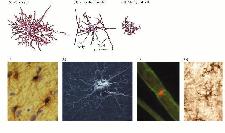
There are three types of glial cells in the mature central nervous system: astrocytes, oligodendrocytes, and microglial cells .Astrocytes, which are restricted to the brain and spinal cord, have elaborate local processes that give these cells a starlike appearance (hence the prefix “astro”). The major function of astrocytes is to maintain, in a variety of ways, an appropriate chemical environment for neuronal signaling. Oligodendrocytes, which are also restricted to the central nervous system, lay down a laminated, lipid-rich wrapping called myelin around some, but not all, axons. Myelin has important effects on the speed of action potential conduction . In the peripheral nervous system, the cells that elaborate myelin are called Schwann cells. As the name implies, microglial cells are smaller cells derived from hematopoietic stem cells (although some may be derived directly from neural stem cells). They share many properties with tissue macrophages, and are primarily scavenger cells that remove cellular debris from sites of injury or normal cell turnover. Indeed, some neurobiologists prefer to categorize microglia as a type of macrophage. Following brain damage, the number of microglia at the site of injury increases dramatically. Some of these cells proliferate from microglia resident in the brain, while others come from macrophages that migrate to the injured area from the circulation.
Nenhum comentário:
Postar um comentário
Observação: somente um membro deste blog pode postar um comentário.