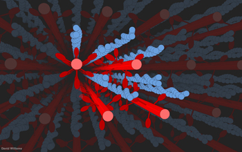
A 3-D computer model of filaments of myosin (in red) reaching out and tugging along filaments of actin (in blue, looking like stands of pearls twined together) during the contraction of a muscle. The model, created by C. David Williams as part of his research into force regulation and the length-tension relationship in muscle, enables researchers to consider the geometry and physics at work on the filaments when a muscle bulges. Williams earned his doctorate at the University of Washington (UW) while conducting this research and is currently a postdoctorate at Harvard University.
This visualization let's researchers see how the individual motor proteins generating force interact with each other to regulate the overall level of force the system develops. The 3-D nature of such models also allows researchers to investigate how the spatial arrangement of a muscles contractile filaments alter the force generated as the muscle goes from very long to very short lengths.
The work was supported by the National Institutes of Health and the National Science Foundation (grant IOS 10-22471, awarded to T.L. Daniel and T.I. Irving to support non-modeling aspects of this research) and with cloud computer access provided by an Amazon.com grant for research.
To learn more, see the UW news story Biceps bulge, calves curve, 50-year-old assumptions muscled aside. (Date of Image: May 2013)
Nenhum comentário:
Postar um comentário
Observação: somente um membro deste blog pode postar um comentário.