Mostrando postagens com marcador Immune system. Mostrar todas as postagens

quarta-feira, 7 de outubro de 2015

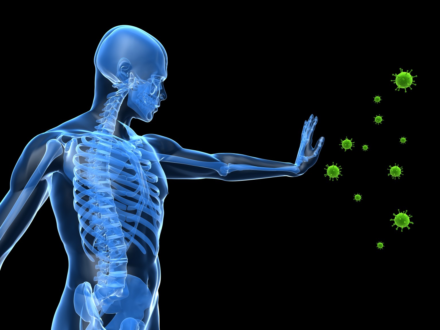
The Science Behind Consciously Controlling Your Immune System: The Mind-Body Connection


The results of a study conducted with the use of Wim Hof method suggest that a person can learn to consciously control his immune responses.

Wim Hof is a Dutch world record holder who is famous worldwide for his ability to resist cold. For this incredible invulnerability to cold, he was commonly nicknamed “the Iceman”.

Scientists led by Matthijs Kox of the Radboud University Medical Center in Nijmegen, the Netherlands, studied his method, which is somehow similar to the Tibetan Tummo technique (yoga of inner heat) and involves third-eye meditation, breathing exercises and cold exposure, and used it to train 12 volunteers to fend off inflammation.

In the framework of the study, 24 volunteers, including 12 people trained in the Wim Hof method and 12 who did not undergo any training, were subjected tothe inflammation test, during which they were injected with a strain of bacteria that provokes flu-like symptoms.

As a result, volunteers who underwent training with Hof method reported fewer and less intense flu-like symptoms than those who did not. At the same time, trained volunteers produced smaller amounts of proteins related to inflammation, and higher levels of interleukin-10, an inflammation-fighting protein.

Hitherto, both the autonomic nervous system and innate immune system were regarded as systems that cannot be voluntarily influenced. The present study demonstrates that, through practicing techniques learned in a short-term training program, the sympathetic nervous system and immune system can indeed be voluntarily influenced,” writes the scientific journal PNAS, where the study was published.

Orthodox neurobiologists and orthodox immunologists have been skeptical. They think the study of the interactions between the nervous and immune systems is a ‘field in the shadows’. This study is a nice way to show that link,” said Guiseppe Matarese, an immunologist of the University of Salerno in Italy who was not involved in the study.

Yet, it is not the only scientific study to show that the mind can control the immune system. In particular, Australian scientists came to the conclusion thatconsciousness is able to influence the intensity of the allergic reaction.

As a part of an experiment, several healthy volunteers had their arms injected with histamine, a substance that plays a major role in many allergic reactions. The experiment was structured so that they thought that the drug was injected in a rubber doll, while a real injection was made in their other arm.

After the experiment, the intensity of the allergic response on both arms of the volunteers was evaluated and compared to the one of the control group who had no rubber doll illusion. As a result, the researchers found that the volunteers who thought that the injection had been given to the doll manifested a much stronger effect of histamine on the arm that had been replaced by the fake one.

“Such a finding is particularly relevant to the immune system because a primary role of the immune system is to discriminate self from non-self,” said Prof Lorimer Moseley, who led the study. “These findings strengthen the argument that the brain exerts some kind of control over specific body parts according to how strongly we own them”.

Both studies show that the immune system can be influenced by the consciousness in many ways, which could mean that our thoughts, beliefs and mood can affect the physical state of our body. Knowing that, you can learn to control your thinking and thus your immune system and health. Focus on the bright side of life, visualize yourself being happy and healthy, meditate every day, and soon you will see positive changes not only in your health but in your whole life!


ABOUT THE AUTHOR

Anna LeMind is the owner and lead editor of the website Learning-mind.com, and a staff writer for The Mind Unleashed.


http://themindunleashed.org/2014/07/science-behind-consciously-controlling-your-immune-system-mind-body-connection.html

quinta-feira, 11 de junho de 2015

Synthetic immune organ produces antibodies


Thu, 06/11/2015 - 9:32am

Anne Ju, Cornell University.

When exposed to a foreign agent, such as an immunogenic protein, B cells in lymphoid organs undergo germinal center reactions. The image on the left is an immunized mouse spleen with activated B cells (brown) that produce antibodies. At right, top: a scanning electron micrograph of porous synthetic immune organs that enable rapid proliferation and activation of B cells into antibody-producing cells. At right, bottom: primary B cell viability and distribution is visible 24 hrs following encapsulation procedure. Images: Singh lab

When exposed to a foreign agent, such as an immunogenic protein, B cells in lymphoid organs undergo germinal center reactions. The image on the left is an immunized mouse spleen with activated B cells (brown) that produce antibodies. At right, top: a scanning electron micrograph of porous synthetic immune organs that enable rapid proliferation and activation of B cells into antibody-producing cells. At right, bottom: primary B cell viability and distribution is visible 24 hrs following encapsulation procedure. Images: Singh labCornell Univ. engineers have created a functional, synthetic immune organ that produces antibodies and can be controlled in the lab, completely separate from a living organism. The engineered organ has implications for everything from rapid production of immune therapies to new frontiers in cancer or infectious disease research.

The immune organoid was created in the lab of Ankur Singh, assistant professor of mechanical and aerospace engineering, who applies engineering principles to the study and manipulation of the human immune system. The work was published online in Biomaterials and will appear later in print.

The synthetic organ is bio-inspired by secondary immune organs like the lymph node or spleen. It is made from gelatin-based biomaterials reinforced with nanoparticles and seeded with cells, and it mimics the anatomical microenvironment of lymphoid tissue. Like a real organ, the organoid converts B cells—which make antibodies that respond to infectious invaders—into germinal centers, which are clusters of B cells that activate, mature and mutate their antibody genes when the body is under attack. Germinal centers are a sign of infection and are not present in healthy immune organs.

The engineers have demonstrated how they can control this immune response in the organ and tune how quickly the B cells proliferate, get activated and change their antibody types. According to their paper, their 3-D organ outperforms existing 2-D cultures and can produce activated B cells up to 100 times faster.

The immune organ, made of a hydrogel, is a soft, nanocomposite biomaterial. The engineers reinforced the material with silicate nanoparticles to keep the structure from melting at the physiologically relevant temperature of 98.6 degrees.

The organ could lead to increased understanding of B cell functions, an area of study that typically relies on animal models to observe how the cells develop and mature.

What’s more, Singh said, the organ could be used to study specific infections and how the body produces antibodies to fight those infections—from Ebola to HIV.

“You can use our system to force the production of immunotherapeutics at much faster rates,” he said. Such a system also could be used to test toxic chemicals and environmental factors that contribute to infections or organ malfunctions.

The process of B cells becoming germinal centers is not well understood, and in fact, when the body makes mistakes in the genetic rearrangement related to this process, blood cancer can result.

“In the long run, we anticipate that the ability to drive immune reaction ex vivo at controllable rates grants us the ability to reproduce immunological events with tunable parameters for better mechanistic understanding of B cell development and generation of B cell tumors, as well as screening and translation of new classes of drugs,” Singh said.

Source: Cornell University

sexta-feira, 17 de abril de 2015

Scientists discover protein that boosts immunity to viruses and cancer

April 16, 2015

Imperial College London

Scientists have discovered a protein that plays a central role in promoting immunity to viruses and cancer, opening the door to new therapies. Experiments in mice and human cells have shown that the protein promotes the proliferation of cytotoxic T cells, which kill cancer cells and cells infected with viruses. The discovery was unexpected because the new protein had no known function and doesn’t resemble any other protein.


Scientists have discovered a protein that plays a central role in promoting immunity to viruses and cancer, opening the door to new therapies.

Experiments in mice and human cells have shown that the protein promotes the proliferation of cytotoxic T cells, which kill cancer cells and cells infected with viruses. The discovery was unexpected because the new protein had no known function and doesn’t resemble any other protein.

Researchers from Imperial College London who led the study are now developing a gene therapy designed to boost the infection-fighting cells, and hope to begin human trials in three years. 

The study also involved researchers at Queen Mary University of London, ETH Zurich and Harvard Medical School. Their discovery, which has been six years in the making, is reported today in the journal Science.

Cytotoxic T cells are an important component of the immune system, but when faced with serious infections or advanced cancer, they are often unable to proliferate in large enough quantities to fight the disease.

By screening mice with genetic mutations, the Imperial team discovered a strain of mice that produced 10 times as many cytotoxic T cells when infected with a virus compared with normal mice. These mice suppressed the infection more effectively, and were more resistant to cancer. They also produced more of a second type of T cells, memory cells, enabling them to recognise infections they have encountered previously and launch a rapid response.

The mice with enhanced immunity produced high levels of a hitherto unknown protein, which the researchers named lymphocyte expansion molecule, or LEM. They went on to show that LEM modulates the proliferation of human T cells as well as in mice.

The researchers now aim to develop a gene therapy designed to improve immunity by boosting the production of LEM. With the support of Imperial Innovations, the technology commercialisation company for the College, the researchers have filed two patents. A company called ImmunarT has been formed with the aim of commercialising the technology.

Professor Philip Ashton-Rickardt from the Section of Immunobiology in the Department of Medicine at Imperial, who led the study, said: “Cancer cells have ways to suppress T cell activity, helping them to escape the immune system. Genetically engineering T cells to augment their ability to fight cancer has been a goal for some time and techniques for modifying them already exist. By introducing an active version of the LEM gene into the T cells of cancer patients, we hope we can provide a robust treatment for patients. 

“Next we will test the therapy in mice, make sure it is safe and see if it can be combined with other therapies. If all goes well, we hope to be ready to carry out human trials in about three years.”

Dr Claudio Mauro, who led the research from the Centre for Biochemical Pharmacology, based within Queen Mary University of London’s William Harvey Research Institute, said: “This study has identified the novel protein LEM and unlocked an unexpected way of enhancing the ability of our immune system to fight viruses or cancers. This is based on the ability of the protein LEM to regulate specific energy circuits, and particularly mitochondrial respiration, in a subset of white blood cells known as cytotoxic T cells. This discovery has immediate consequences for the delivery of innovative therapeutic approaches to cancer. Its ramifications, however, are far greater as they can help explaining the biological mechanisms of widespread human diseases involving altered immune and inflammatory responses. These include chronic inflammatory and autoimmune disorders, such as atherosclerosis and rheumatoid arthritis.”

The research was funded by the Medical Research Council, the Wellcome Trust and the British Heart Foundation.

Dr Mike Turner, Head of Infection and Immunobiology at The Wellcome Trust, said: “The discovery of a protein that could boost the immune response to not only cancer, but also to viruses, is a fascinating one. Further investigation in animal models is needed before human trials can commence, but there is potential for a new type of treatment that capitalises on the immune system’s innate ability to detect and kill abnormal cells.”

terça-feira, 14 de abril de 2015

Recruiting the entire immune system to attack cancer

Tue, 04/14/2015 - 11:30am

Anne Trafton, MIT News Office

The human immune system is poised to spring into action at the first sign of a foreign invader, but it often fails to eliminate tumors that arise from the body’s own cells. Cancer biologists hope to harness that untapped power using an approach known as cancer immunotherapy.

Orchestrating a successful immune attack against tumors has proven difficult so far, but a new study from Massachusetts Institute of Technology (MIT) suggests that such therapies could be improved by simultaneously activating both arms of the immune system. Until now, most researchers have focused on one of two strategies: attacking tumors with antibodies, which activate the innate immune system, or stimulating T cells, which form the backbone of the adaptive immune system.

By combining these approaches, the MIT team was able to halt the growth of a very aggressive form of melanoma in mice.

“An anti-tumor antibody can improve adoptive T-cell therapy to a surprising extent,” says Dane Wittrup, the Carbon P. Dubbs Professor in Chemical Engineering at MIT. “These two different parts of the immune therapy are interdependent and synergistic.”

Wittrup, an associate director of MIT’s Koch Institute for Integrative Cancer Research and also a faculty member in the Department of Biological Engineering, is the senior author of a paper describing the work in Cancer Cell. Lead authors are graduate students Eric Zhu and Cary Opel and recent PhD recipient Shuning Gai.

Enlisting the immune system
Antibody drugs for cancer, which include rituximab and Herceptin, are believed to work by binding to cancer proteins and blocking the signals that tell cancer cells to divide uncontrollably. They may also draw the attention of cells belonging to the innate immune system, such as natural killer cells, which can destroy tumor cells.

Adoptive T cell therapy, on the other hand, enlists the body’s T cells to attack tumors. Billions of T cells flow through the average person’s bloodstream at any given time, each specialized to recognize different molecules. However, many tumor proteins do not provoke T cells to attack, so T cells must be removed from the patient and programmed to attack a specific tumor molecule.

Wittrup and his colleagues made the discovery that they could generate both types of immune responses while they were experimenting with improving antibody drug performance with a signaling molecule called IL-2, which helps boost immune responses.

Scientists have tried this strategy before, and about a dozen such therapies have gone through phase I clinical trials. However, most of these efforts failed, even though the antibody-IL-2 combination usually works very well against cancer cells grown in a lab dish.

The MIT team realized that this failure might be caused by the timing of IL-2 delivery. When delivered to cells in a dish, IL-2 sticks around for a long time, amplifying the response of natural killer cells against cancer cells. However, when IL-2 is injected into a patient’s bloodstream, the kidneys filter it out within an hour.

Wittrup and his colleagues overcame this by fusing IL-2 to part of an antibody molecule, which allows it to circulate in the bloodstream for much longer. In tests in mice with a very aggressive form of melanoma, the researchers found they could stop tumor growth by delivering this engineered form of IL-2, along with antibody drugs, once a week.

Immune synergy
To their surprise, the researchers found that T cells were the most important component of the anti-tumor response induced by the antibody-IL-2 combination. They believe that the synergy of IL-2-induced cells and cytokines, and the antibody treatment, creates an environment that lets T cells attack more effectively.

“The antibody-driven innate response creates an environment such that when the T cells come in, they can kill the tumor. In its absence, the tumor cells establish an environment where the T cells don’t work very well,” Wittrup says.

Cells called neutrophils, which are considered the immune system’s “first line of defense” because they react strongly to foreign invaders that enter the skin through a cut or other injury, were also surprisingly important.

“They’re a really powerful force in your immune system, but people in immunotherapy don’t usually focus on neutrophils. They don’t really consider them as a viable tool,” Zhu says. “It pointed us to the idea that although T cells and natural killer cells are important, maybe we’re forgetting about a part of the immune system that is also really important and could help us achieve our goals of ultimately curing the tumors.”

The researchers also found that when they delivered an antibody, IL-2, and T cells targeted to the tumor, the adoptively transferred T cells killed cancer cells much more successfully than when only T cells were delivered. In 80 to 90% of the mice, tumors disappeared completely; even when tumor cells were re-injected into the mice months after the original treatment, their immune systems destroyed the cells, preventing new tumors from forming

In a related paper that appeared recently in the Proceedings of the National Academy of Science, the MIT team also found that delivering IL-2 bound to any kind of antibody, even if the antibody did not target a protein on the tumor cell surface, would halt or slow tumor growth, especially if additional doses of the antibody alone were also given. Graduate student Alice Tzeng was the lead author of that study.

The researchers are now exploring additional proteins that could be added to the IL-2 and antibody combination to make immunotherapy more effective. In the meantime, simply giving patients more prolonged exposure to IL-2 could improve the effectiveness of existing antibody drugs, Wittrup says.

Source: Massachusetts Institute of Technology

ENTRIES OPEN:
Establish your company as a technology leader. For 50 years, the R&D 100 Awards, widely recognized as the “Oscars of Invention,” have showcased products of technological significance. Learn more.

sábado, 14 de fevereiro de 2015

New mechanism that controls immune responses discovered

UT Southwestern Medical Center researchers have identified a common signaling mechanism to produce interferon -- one of the main proteins used to signal the immune system when the body needs to defend itself against a virus, tumor, or other diseases.

The findings are important for understanding the body's immune defense system, searching for compounds to turn the immune system on or off, and they may help combat autoimmune diseases, in which overactive immune cells attack healthy tissues.

"Our work reveals a common mechanism by which three distinct pathways lead to the production of type-I interferons," said Dr. Zhijian "James" Chen, Professor of Molecular Biology and in the Center for the Genetics of Host Defense at UT Southwestern, and a Howard Hughes Medical Institute (HHMI) Investigator. "Ultimately, we believe that understanding this mechanism will facilitate the design and development of medications to treat human diseases such as lupus."

The findings appear online in the journal Science.

The results show how a protein called interferon regulatory factor 3 (IRF3), which controls the production of type-I interferons, is activated and how this pathway is tightly controlled. The failure of this control system can lead to autoimmune disorders such as systemic lupus erythematosus, which causes joint pain and swelling, and can damage the brain, heart, lungs, kidneys, and digestive track. Lupus affects more than 1.5 million Americans, and is more common in young and middle-aged women than in men.

A normal function of interferons is to defend the body against infections from viruses, bacteria and parasites. Previous research has identified specific pathways that induce interferons in response to distinct infectious agents, but how these different pathways converge on IRF3 to induce interferons was not understood.

Dr. Chen and his team studied a protein called MAVS, which they discovered in 2005 and showed that it is an adaptor protein essential for interferon induction by RNA viruses such as influenza virus. In the new study, they found that MAVS is modified by the addition of a phosphate group (phosphorylated) by an enzyme called TBK1 when cells are infected by a virus and that this modification is important for IRF3 activation.

Upon closer examination, they found the amino acid sequence that is phosphorylated in MAVS is very similar to those of two other adaptor proteins, STING and TRIF, which mediate interferon induction in response to DNA viruses and bacteria, respectively. Further research confirmed that all three adaptor proteins are phosphorylated at the common sequence motif and that this phosphorylation allows each of the adaptor proteins to bind IRF3, thereby facilitating IRF3 phosphorylation by TBK1. The phosphorylated IRF3 becomes activated to induce type-I interferons.

"Although TBK1 is required for IRF3 activation, TBK1 alone is not sufficient. Phosphorylation of the adaptor proteins provides a 'license' for TBK1 to phosphorylate IRF3," said Dr. Chen, who holds the George L. MacGregor Distinguished Chair in Biomedical Science. "This hitherto unrecognized mechanism ensures that type-I interferons are produced only when a proper adaptor protein is engaged in cells that are infected by pathogens."


Story Source:

The above story is based on materials provided by UT Southwestern Medical Center. Note: Materials may be edited for content and length.


Journal Reference:

  1. S. Liu, X. Cai, J. Wu, Q. Cong, X. Chen, T. Li, F. Du, J. Ren, Y. Wu, N. Grishin, Z. J. Chen. Phosphorylation of innate immune adaptor proteins MAVS, STING, and TRIF induces IRF3 activation. Science, 2015; DOI: 10.1126/science.aaa2630


segunda-feira, 9 de fevereiro de 2015

A Pill That Mimics the Immune System


Synthetic antibodies that can be orally ingested to fight cancer and autoimmune diseases may replace cumbersome intravenous therapeutics

February 9, 2015 By Bret Stetka

antibodies

The therapeutic potential of synthetic antibodylike compounds is vast.
Credit: NIAID via Flickr

The human body doesn’t like outsiders. When a foreign pathogen or substance, say an unwanted virus, finds its way into our blood streams we produce antibodies that the neutralize the threat. These “Y”-shaped proteins are made by a class of white blood cells called plasma cells and bind to molecules on the invaders called antigens, triggering another set of white blood cells to literally ingest the interloper. For years now doctors have used antibodies and other protein-based therapies (aka biologics) to treat a range of illnesses, cancers, infections and autoimmune diseases among them. There’s only one problem: antibodies are bulky and hence usually have to be administered intravenously as they’re often to big to be absorbed in the gastrointestinal tract. With this in mind, chemist David Spiegel and his colleagues at Yale University are out to simplify dosing for patients on antibody therapy by developing treatments with the benefits of antibodies—minus the needle.

In work recently published in the Journal of the American Chemical Society Spiegel and his team have successfully developed the first synthetic molecules that behave like antibodies. Like the real thing, these so-called "synthetic antibody mimics"—or "SyAMs"—bind to both diseased cells and disease-fighting immune cells. Specifically the compounds were found to zero in on and bind to a specific antigen on prostate cancer cells. The SyAMs also bind to and activate certain immune cells that then devour the malignancy.

Spiegel’s SyAMs are produced in a way that is similar to conventional drugs, by using chemical reactions to piece together various structural features often not found in nature. As he explains, the therapeutic potential of synthetic antibodylike compounds is vast: “Because antibodies are proteins they’re difficult and expensive to produce on a large scale, can cause unwanted immune reactions and tend to aggregate and denature with long-term storage.” Spiegel speculates that SyAMs will be easier and cheaper to produce and less likely to incite aberrant immune activity. SyAMs are also one twentieth the size of antibodies—more akin to the size of most medications—and can therefore perhaps be administered orally. This could be a major boon to patients with cancers and autoimmune diseases like multiple sclerosis who have to regularly get themselves to infusion centers for monoclonal antibody therapy.

The idea of producing “artificial antibodies” traces back to the work of late 19th-century German physician Paul Ehrlich who first proposed that the immune system can neutralize toxins or pathogens by forming "antitoxins." Based on the idea he and his colleagues began developing drugs meant to function like these antitoxins, including one to treat Trypanosoma parasite infections. During the 1930s and 1940s—as understanding of antibody–antigen interactions grew—famed chemist, activist and vitamin C evangelist Linus Pauling started tinkering with the idea that proteins could be transformed into antibodies by exposing them to certain antigens.

Artificial antibody research split subsequently in two directions: one camp pursued creating protein antibodies resulting in what are called monoclonal antibodies. Monoclonals are produced by natural means in a lab and are now commonly used therapeutically. The other camp, in which Spiegel falls, set out to produce smaller, nonprotein compounds with antibodylike properties.

Beyond attacking prostate cancer, Spiegel’s group has also developed SyAM-based approaches targeting HIV, various other cancers and bacterial triggers of autoimmune disease. And although SyAM research remains in the petri dish, a mouse model is in the works and human studies are not far off. A number of other labs are also researching ways to fight disease by manipulating antibodies and synthesizing molecules that act on the immune system, including Peter Schultz at the Scripps Research Institute in La Jolla, Calif. “He's probably our biggest competitor and I'm his biggest fan,” Spiegel says.

Laura Kiessling at the University of Wisconsin–Madison, who studies ways to draw natural antibodies to tumor cells, comments on the benefits of Spiegel’s approach: “It can be tailored to selectively recruit specific types of immune cells to kill tumor cells. The smaller size of the compounds could also be an asset in eliminating tumors, but the benefits would need to be looked at in vivo,” Kiessling says.

As Spiegel confesses, his route to the chemistry lab was an unusual one. He went to medical school to become a psychiatrist but along the way also picked up an interest in chemistry and—in decidedly un-psychiatristlike fashion—tacked on a PhD in organic synthesis (the development of new organic compounds and reactions in the lab). “The relevance of this research to clinical medicine was not always clear to my classmates and colleagues—or sometimes to myself,” he recalls, “but I realized that organic chemistry has been critically associated with drug development from the beginning.”

Spiegel points out that most U.S. Food and Drug Administration–approved drugs are in fact small organic molecules. “My thought was that by working to understand diseases at the cellular and molecular level, I could not only learn how to make new drugs,” he notes, “but also perhaps develop new paradigms for how drugs could function.” Spiegel’s decision to join his chemical and clinical interests, it seems, was a wise one.

source : www.scientificamerican.com

sexta-feira, 16 de janeiro de 2015

Tumor suppressor protein plays key role in maintaining immune balance


A new study from St. Jude Children’s Research Hospital shows that the PTEN tumor suppressor protein is essential for proper regulatory T cell function. The discovery offers new focus for improving treatment of autoimmune diseases. Corresponding author is Hongbo Chi, Ph.D. (on right).

St. Jude Children’s Research Hospital scientists have discovered that a protein widely known for suppressing tumor formation also helps prevent autoimmune diseases and other problems by putting the brakes on the immune response. The research was published recently online ahead of print in the scientific journal Nature Immunology.

Researchers showed that the tumor suppressor protein PTEN is essential for proper functioning of regulatory T cells. This small population of white blood cells helps to maintain immune system balance by suppressing specialized T cells called helper T cells that fuel distinct parts of the immune response. The helper T cells investigated in this study included type 1 T helper (Th1) and follicular T helper (Tfh) cells.

The interplay between regulatory T cells and helper T cells is crucial for both combating infections and for preventing misguided immune attacks that lead to autoimmune diseases and other problems. But details of how regulatory T cells control the diverse functions of various helper T cells have been elusive. This study fills key gaps in that understanding, particularly PTEN’s role. The work also identified a new focus for research to improve treatment of autoimmune diseases.

PTEN is best known as one of the most frequently altered tumor suppressor genes in human cancers, but loss of the protein has also been tied to autoimmune problems. This study showed that is because PTEN is required to maintain the stable population of regulatory T cells that keeps the immune system in check.

“In humans we know that loss of PTEN leads to tumors. This study highlights another role and shows that PTEN is also crucial for proper functioning of regulatory T cells and prevention of autoimmune diseases,” said corresponding author Hongbo Chi, Ph.D., a member of the St. Jude Department of Immunology. “In mice, the loss of just one copy of the PTEN gene in regulatory T cells is sufficient to set the stage for autoimmune problems.”

Working in specially bred mice, researchers showed that deleting the PTEN gene in regulatory T cells was followed by a dramatic increase in the number of Tfh and related cells. Tfh cells aid production of antibodies, which combat infections. But when produced inappropriately, antibodies can also drive autoimmune disorders like lupus. The mice in this study developed kidney damage and immune changes associated with lupus. Restoring PTEN to 50 percent of normal levels did not protect the mice from inflammatory disease.

Researchers found evidence that Th1 cells influence the activity of Tfh cells. Th1 cells produce the chemical messenger interferon gamma that revs up the immune response. When researchers blocked interferon gamma production in the specially bred mice, the number of Tfh cells fell along with lupus-like immune abnormalities.

“We have identified a crucial role of PTEN in controlling Tfh cells and autoantibody production. Additionally, by linking the role of PTEN to Tfh cells, we have opened doors for further investigation of Tfh related lymphomas,” said co-first author Sharad Shrestha, a graduate student in Chi’s laboratory. Added co-first author Kai Yang, Ph.D., a staff scientist in Chi’s laboratory: “These results reveal a hierarchy of control that regulatory T cells use to simultaneously regulate Th1 and Tfh cells. We showed that Th1 production of interferon gamma is a pre-requisite for the activity of Tfh cells.”

The findings also yielded insight into a cell signaling pathway that regulates many important functions, including T cell activity, in response to changing conditions. This is the mTOR pathway, in which the protein complexes mTORC1 and mTORC2 play central roles.

Investigators showed that deletion of PTEN in regulatory T cells led to increased activity of mTORC2 but not mTORC1. When scientists blocked mTORC2 activity in mice whose regulatory T cells lacked PTEN, immune system balance and activity returned to normal. “Our research establishes that the interaction of PTEN and mTORC2 functions as a central pathway to maintain the stability of the regulatory T cell population and to ensure their ability to control the activity of Th1 and Tfh cells,” Chi said. The newly identified PTEN-mTORC2 axis provides another target for efforts to develop better treatments of autoimmune and other disorders.

sexta-feira, 12 de dezembro de 2014

Getting antibodies into shape to fight cancer


December 11, 2014

University of Southampton

The precise shape of an antibody makes a big difference to how it can stimulate the body's immune system to fight cancer, paving the way for much more effective treatments, researchers have found. The latest types of treatment for cancer are designed to switch on the immune system, allowing the patient's own immune cells to attack and kill cancerous cells, when normally the immune cells would lie dormant. In a new study, a research team has found that a particular form of antibody, called IgG2B, is much more effective at stimulating cancer immunity than other types.


Scientists at the University of Southampton have found that the precise shape of an antibody makes a big difference to how it can stimulate the body's immune system to fight cancer, paving the way for much more effective treatments.

The latest types of treatment for cancer are designed to switch on the immune system, allowing the patient's own immune cells to attack and kill cancerous cells, when normally the immune cells would lie dormant.

In a study, funded by Cancer Research UK and published in the journal Cancer Cell, the Southampton team have found that a particular form of antibody, called IgG2B, is much more effective at stimulating cancer immunity than other types. Unlike other forms of antibody, IgG2B can work independently without needing help from other immune cells, making it more active and able to work in all tissues of the body. The team have also been able to engineer antibodies that will be locked into the particular shape (called a locked B structure) that is most active, making them much stronger immune stimulators than previous drugs.

Dr Ann White, who led the study, comments: "We know that the immune system provides a natural protection against cancer, which can only grow by finding a way around our defences. Antibody treatments are now able to correct this problem for many types of cancer, but we still need them to work better..

"It is early days, but this important discovery could enable us to treat more cancers effectively. Our next task is to bring these novel IgG2B antibodies into trials for cancer patients and we are engineering ways to make them effective in the clinic."

The team is now working to discover why the IgG2 molecule works better in a locked B structure. To do this the molecule has been crystallised and an x-ray shone through it so the structure can be assessed. It is the first time that the IgG2 has been crystallised.

Professor Nic Jones, Cancer Research UK's chief scientist, said: "This research has zeroed in on how we can make immunotherapy treatments more effective against cancer. Energising the immune cells in our body and getting them to treat cancer cells as a threat gives us a better shot at beating cancer.. Immunotherapy is part of the future of cancer treatment and it's important that we use our best immunotherapy weapons to fight the disease."


Story Source:

The above story is based on materials provided by University of Southampton. Note: Materials may be edited for content and length.


Journal Reference:

  1. Ann L. White, H.T. Claude Chan, Ruth R. French, Jane Willoughby, C. Ian Mockridge, Ali Roghanian, Christine A. Penfold, Steven G. Booth, Ali Dodhy, Marta E. Polak, Elizabeth A. Potter, Michael R. Ardern-Jones, J. Sjef Verbeek, Peter W.M. Johnson, Aymen Al-Shamkhani, Mark S. Cragg, Stephen A. Beers, Martin J. Glennie. Conformation of the Human Immunoglobulin G2 Hinge Imparts Superagonistic Properties to Immunostimulatory Anticancer Antibodies. Cancer Cell, 2014; DOI: 10.1016/j.ccell.2014.11.001


quarta-feira, 26 de novembro de 2014

Selenium compounds boost immune system to fight against cancer


Cancer types such as melanoma, prostate cancer and certain types of leukemia weaken the body by over-activating the natural immune system. Researchers from the University of Copenhagen have now demonstrated that selenium -- naturally found in, e.g., garlic and broccoli -- slows down the immune over-response. In the long term, this may improve cancer treatment. The findings have been published in the Journal of Biological Chemistry.

The immune system is designed to remove things not normally found in the body. Cells undergoing change, e.g. precursors of cancer cells, are therefore normally recognised and removed by the immune system. Unfortunately, the different cancer cells contain mechanisms that block the immune system's ability to recognise them, allowing them to freely continue cancer development.

Certain cancer cells overexpress immunostimulatory molecules in liquid form. Such over-stimulation has a negative impact on the immune system:

"You can say that the stimulating molecules over-activate the immune system and cause it to collapse, and we are, of course, interested in blocking this mechanism. We have now shown that certain selenium compounds, which are naturally found in, e.g., garlic and broccoli, effectively block the special immunostimulatory molecule that plays a serious role for aggressive cancers such as melanoma, prostate cancer and certain types of leukemia," says Professor Søren Skov, Department of Veterinary Disease Biology, University of Copenhagen.

Dissolved molecules

In this study, the researchers are focusing on the so-called NGK2D ligands. There are eight variants, of which one in particular has caught the researchers' attention, because it assumes liquid form. It is precisely the molecular dissolution that causes serious problems, once the cancer is raging. The entire bloodstream is, so to speak, infected, and the molecule is therefore used as a marker of serious illness:

"Molecules are found both on the surface of the cancer cells and dissolved in the blood of the affected person. We are now able to show that selenium compounds appear to have a very beneficial effect when it comes to neutralising the special variant of the NGK2D ligand -- both in soluble form and when the molecule is placed on the cell surface," says Professor Søren Skov.

Better drugs in future

The researchers are constantly learning more about the disease mechanisms causing aggressive cancers in the skin, blood and reproductive organs:

"The overexpression seen in cancers such as melanoma, prostate cancer and certain types of leukemia significantly impairs the immune system. If we can find ways to slow down the over-stimulation, we are on the right track. The new results are yet another small step towards better cancer drugs with fewer adverse effects," says Søren Skov.


Story Source:

The above story is based on materials provided by University of Copenhagen - The Faculty of Health and Medical Sciences. Note: Materials may be edited for content and length.


Journal Reference:

  1. M. Hagemann-Jensen, F. Uhlenbrock, S. Kehlet, L. Andresen, C. Gabel-Jensen, L. Ellgaard, B. Gammelgaard, S. Skov. The Selenium Metabolite Methylselenol Regulates the Expression of Ligands That Trigger Immune Activation through the Lymphocyte Receptor NKG2D. Journal of Biological Chemistry, 2014; 289 (45): 31576 DOI: 10.1074/jbc.M114.591537


sábado, 8 de novembro de 2014

A cause of age-related inflammation found


November 6, 2014

Carnegie Institution

As animals age, their immune systems gradually deteriorate, a process called immunosenescence. It is associated with systemic inflammation and chronic inflammatory disorders, as well as with many cancers. The causes underlying this age-associated inflammation, and how it leads to diseases, are poorly understood. New work sheds light on one protein's involvement in suppressing immune responses in aging fruit flies.


This image shows a comparison of lamin-B in the fat bodies of 10-day-old and 50-day-old fruit flies.

As animals age, their immune systems gradually deteriorate, a process called immunosenescence. It is associated with systemic inflammation and chronic inflammatory disorders, as well as with many cancers. The causes underlying this age-associated inflammation, and how it leads to diseases, are poorly understood. New work in Carnegie's Yixian Zheng's lab sheds light on one protein's involvement in suppressing immune responses in aging fruit flies. It is published in Cell.

Insects have an immune organ called the fat body, which is roughly equivalent to the mammalian fat and liver. It is responsible for many immune functions. Zheng and her team--Carnegie's Haiyang Chen and Xiaobin Zheng--found that the fruit fly fat body experiences a great deal of inflammation in aged flies.

These inflamed fly fat bodies then secrete proteins that lead to a reduction in immune response of the gut. This reduction of the gut immune response causes the gut's stem cells to undergo excessive division and inappropriate differentiation, creating a condition called hyperplasia that shares features with the precancerous polyps found in human guts.

Zheng and her team found that the gradual reduction of a protein called lamin-B in the fat bodies of aging flies is the culprit behind fat body inflammation and the resulting hyperplastic gut, all of which falls under the umbrella of immunosenescence.

Lamin-B is part of the lamin family of proteins, which form the major structural component of the material that lines the inside of a cell's nucleus. Lamins have diverse functions, including suppressing gene expression, and they are found in an array of tissues and organs. In humans, diseases caused by mutations in lamins are called laminopathies and include premature aging.

B-type lamins have long been suspected to play a role in gene suppression by binding to segments of DNA. The team's work revealed that when the fruit fly fat body was depleted of lamin-B, the normal suppression of genes involved in the immune response is reversed, just as it would be in response to bacterial infection or injury, but in this case there is no apparent infection or injury. The un-suppressed immune response initiates the inflammation and resulting gut hyperplasia.

"Our findings have implications for mammals as well as for insects, as immune response genes in mammals also are known to have lamins present on them," Zheng explained. "We think that lamin-B might play an evolutionarily conserved role in suppressing inflammatory genes in immune organs in the absence of infection or injury and our work could provide insight into immunosenescence in humans."


Story Source:

The above story is based on materials provided by Carnegie Institution. Note: Materials may be edited for content and length.


Journal Reference:

  1. Haiyang Chen, Xiaobin Zheng, Yixian Zheng. Age-Associated Loss of Lamin-B Leads to Systemic Inflammation and Gut Hyperplasia. Cell, 2014; 159 (4): 829 DOI: 10.1016/j.cell.2014.10.028


quinta-feira, 2 de outubro de 2014

Adolescent exposure to THC may cause immune systems to go up in smoke


When it comes to using marijuana, new research, involving mice and published in the October 2014 issue of the Journal of Leukocyte Biology, suggests that just because you can do it, doesn't mean that you should. That's because a team of Italian scientists have found that using marijuana in adolescence may do serious long-term damage to the immune system. This damage may result in autoimmune diseases and chronic inflammatory diseases, such as multiple sclerosis, inflammatory bowel disease and rheumatoid arthritis in adulthood.

"I hope that the knowledge that early exposure to marijuana is associated with immediate and long-term deleterious effects on the immune system may reach adolescents and their families," said Paola Sacerdote, Ph.D., a researcher involved in the work from the Università degli Studi di Milano in Milano, Italy. "The increased risk of getting sick in adulthood may hopefully be a deterrent for marijuana abuse among young individuals."

To make this discovery, scientists injected "adolescent" mice with THC, the main active component of marijuana, for 10 days. This period in the mouse lifecycle corresponded to the adolescence period in humans (approximately ages 12-18). A second group of adolescent mice received only a placebo. At the end of treatments, both groups of animals were left undisturbed for approximately two months, until they reached full adulthood. The activity of the immune system was then evaluated, taking into consideration several important measurements, such as the ability of leukocytes to produce cytokines to mount an antibody response to vaccination or the capacity of macrophage to phagocyte particles. The group of mice treated with THC in adolescence had severe alterations of immune responses in adulthood, characterized by a clear switch toward a pro-inflammatory and cytotoxic phenotype.

"The immune system is characterized by an impressive ability to 'remember' previous exposures and changes during the period of immune system development especially early in life can have important long-term consequences," said John Wherry, Ph.D., Deputy Editor of the Journal of Leukocyte Biology. "These studies not only point to adolescence as a key phase of immune system sensitivity, but also highly the dramatic and long-lasting negative effects that a common recreational drug abused by teenagers may have on immune function."


Story Source:

The above story is based on materials provided by Federation of American Societies for Experimental Biology. Note: Materials may be edited for content and length.


Journal Reference:

  1. S. Moretti, M. Castelli, S. Franchi, M. A. Raggi, L. Mercolini, M. Protti, L. Somaini, A. E. Panerai, P. Sacerdote. 9-Tetrahydrocannabinol-induced anti-inflammatory responses in adolescent mice switch to proinflammatory in adulthood. Journal of Leukocyte Biology, 2014; 96 (4): 523 DOI: 10.1189/jlb.3HI0713-406RR


domingo, 28 de setembro de 2014

Protecting the body from itself: How defense cells fight disease, but not themselves


Autoimmune B cells cause the over-activation and death of NKT cells via presentation of altered lipids.

Scientists from A*STAR's Bioprocessing Technology Institute (BTI) have established a clearer relationship between two cells which serve our body's natural defence mechanisms against diseases and infections. Their findings, published in the journal CELL REPORTS, will help the medical community better understand autoimmunity and could pave the way for treatment of autoimmune diseases.

Natural killer T (NKT) cells and B cells are two of many immune cell types that work in tandem to help the body fight against foreign infectious agents. NKT cells have very potent functions and are crucial to the immune system despite making up only a small percentage of white blood cells. While scientists have established that NKT cells can promote the production of antibodies by B cells to combat infection, little is known about the effect of B cells on NKT cells until now.

Patients with autoimmune disorders have been observed to have drastically reduced numbers of NKT cells. The study conducted by BTI scientists revealed that autoimmune B cells had altered lipid compositions, causing NKT cells to be over-activated and resulting in their eventual death and depletion in numbers. The scientists further found that removal of a lipid-presenting molecule from B cells resulted in recovery in the numbers of NKT cells.

Dr Andy Tan, a research scientist at BTI who led the study, said, "Our findings provide an alternative theory to current understanding of how autoimmune B cells affect NKT cells. This will allow new therapeutic strategies to be devised, rectifying NKT cell deficiency in autoimmune patients and improving their health."

Autoimmune diseases, including lupus and some forms of arthritis and diabetes, develop when the immune system fails to distinguish between its own cells and foreign pathogens such as bacteria and viruses, resulting in attacks on the patient's own healthy tissue. Autoimmunity affects between three to 10 percent of the general population, depending on gender, disease type and geographical location. Future studies to identify particular lipids responsible for NKT cell over-activation might thus present therapeutic opportunities for those with autoimmune diseases and disorders.

Prof Lam Kong Peng, senior author for the study and Executive Director of BTI, added, "One of the central questions in the field of immunology is how the immune system is able to maintain a delicate balance in effectively fighting foreign pathogens and avoiding attack of the body itself. Our findings have brought us one step closer to unravelling the mechanisms that govern the balance between immunity and autoimmunity."


Story Source:

The above story is based on materials provided by A*Star Agency for Science, Technology and Research. Note: Materials may be edited for content and length.


Journal Reference:

  1. Andy Hee-Meng Tan, William Pooi-Kat Chong, Sze-Wai Ng, Nurhidayah Basri, Shengli Xu, Kong-Peng Lam. Aberrant Presentation of Self-Lipids by Autoimmune B Cells Depletes Peripheral iNKT Cells. Cell Reports, 2014; DOI: 10.1016/j.celrep.2014.08.043


sábado, 27 de setembro de 2014

Researchers contribute to study of trained immunity


September 26, 2014

The Geisel School of Medicine at Dartmouth

Support for a new -- and still controversial -- understanding of the immune system has been released in a new study. Epigenetic profiling experiments identified genes involved in glucose metabolism as being critical for trained immunity. These findings have potential implications both for the prevention and treatment of inflammatory diseases and for bolstering the immune response to pathogens in situations where the immune system is not functioning properly.


A study published in the journal Science provides support for a new -- and still controversial -- understanding of the immune system. The research was conducted by collaborators in the U.S. and Europe, including Robert Cramer, PhD, an assistant professor of microbiology and immunology at the Geisel School of Medicine and member of the Dartmouth Lung Biology Center, and Kelly Shepherdson, PhD, at the time a graduate student in Cramer's lab.

Typically, scientists divide the immune system into two categories: the innate immune response and the adaptive immune response. The adaptive immune response is familiar to most people because of its role in providing long-term protection against disease, as when a vaccine triggers the immune system to "remember" a specific threat and mount a robust response if challenged by that pathogen later in life.

The innate immune system is older in evolutionary terms and usually thought of as responding to immediate threats from pathogens or other foreign entities. But over the past few years, informed by studies in plants and invertebrates, scientists have begun to suspect that the innate response has a form of "memory" as well, complicating the division of the immune system into two neat categories. "The innate immune system is typically thought of as a quick, generally non-specific broad response to an initial infection that lacks immunological memory," says Cramer. "But studies of trained immunity suggest that maybe that's not the case, and that is not only paradigm shifting but potentially directly relevant to the treatment and prevention of many diseases."

In 2011, researchers in the Netherlands, including Mihai Netea, MD, PhD, the senior author on the new paper in Science, coined the term "trained immunity" to refer to immune responses that involve immunological memory deriving from cells associated with the innate immune system. Trained immunity is capable of providing non-specific protection from secondary infections. However, the mechanisms of how trained immunity is initiated and maintained remain unclear. Recent research has found that epigenetic changes occurred in innate immune cells when exposed to certain pathogens or their antigens and are an important feature of trained immunity. But the specific genes and biochemical pathways associated with trained cells were unknown.

In the Science paper, the researchers report that changes in metabolism are a critical driving force behind the trained immunity phenotype. Epigenetic profiling experiments identified genes involved in glucose metabolism as being critical for trained immunity. Cramer and Shepherdson became involved in the research because of their work on HIF1α and the innate immune response to fungi. HIF1α is a protein that acts as a transcription factor for genes involved in metabolism, among other genes.

Using mice in which the HIF1α gene was deleted from cells of the innate immune system, Cramer and Shepherdson tested the hypothesis that changes in metabolism, mediated in part through the HIF1α pathway, were critical for trained immunity. They first exposed the mice to a fungal polysaccharide antigen, beta glucan, that induces a trained immune response and then challenged the mice with a bacterial pathogen that can cause sepsis. The normal mice were protected against the pathogen by the trained immune response, but the mice without HIF1α were not protected, indicating that the lack of HIF1α prevented the trained immune response and protection against secondary infection. "This was a critical set of experiments for the research as they were conducted in vivo in a whole animal that lacked HIF1α in the key effector cells of trained immunity," Cramer says.

These findings have potential implications both for the prevention and treatment of inflammatory diseases and for bolstering the immune response to pathogens in situations where the immune system is not functioning properly. A next step in this line of research, Cramer says, is to identify genes downstream of HIF1α critical for the trained phenotype that may make viable specific targets for therapeutic development. "If we can figure out these underlying mechanisms, we might be able to enhance the efficacy of vaccines," he says. And, he adds, it might also be time to rethink the traditional understanding of the immune system.


Story Source:

The above story is based on materials provided by The Geisel School of Medicine at Dartmouth. The original article was written by Amos Esty. Note: Materials may be edited for content and length.


Journal Reference:

  1. S.-C. Cheng, J. Quintin, R. A. Cramer, K. M. Shepardson, S. Saeed, V. Kumar, E. J. Giamarellos-Bourboulis, J. H. A. Martens, N. A. Rao, A. Aghajanirefah, G. R. Manjeri, Y. Li, D. C. Ifrim, R. J. W. Arts, B. M. J. W. van der Meer, P. M. T. Deen, C. Logie, L. A. O'Neill, P. Willems, F. L. van de Veerdonk, J. W. M. van der Meer, A. Ng, L. A. B. Joosten, C. Wijmenga, H. G. Stunnenberg, R. J. Xavier, M. G. Netea. mTOR- and HIF-1 -mediated aerobic glycolysis as metabolic basis for trained immunity. Science, 2014; 345 (6204): 1250684 DOI: 10.1126/science.1250684


segunda-feira, 22 de setembro de 2014

Immune system of newborn babies stronger than previously thought


The team discovered that whilst T cells in newborn babies are largely different to those in adults, it is not because they are immunosuppressed; rather, they manufacture a potent anti-bacterial molecule known as IL8 that has not previously been considered a major product of T cells, and that activates neutrophils to attack the body's foreign invaders.

Contrary to what was previously thought, newborn immune T cells may have the ability to trigger an inflammatory response to bacteria, according to a new study led by King's College London. Although their immune system works very differently to that of adults, babies may still be able to mount a strong immune defense, finds the study published in the journal Nature Medicine.

Our immune system is made up of several different types of immune cells, including neutrophils which play an important role in the frontline defense against infection, and lymphocytes: B cells which produce antibodies, and T cells that target cells infected with viruses and microbes.

Up to now, it was generally believed that babies have an immature immune system that doesn't trigger the same inflammatory response normally seen in adults. Although babies need to protect themselves from the harmful pathogens they are exposed to from birth, it was thought that their T cells were suppressed to some extent to prevent inflammatory damage to the developing child. Sceptical of this notion, the King's-led study set out to characterize the properties of T cells, examining very small samples of blood in twenty-eight highly premature babies, as they developed over the first few weeks of life.

The team discovered that whilst T cells in newborn babies are largely different to those in adults, it is not because they are immunosuppressed; rather, they manufacture a potent anti-bacterial molecule known as IL8 that has not previously been considered a major product of T cells, and that activates neutrophils to attack the body's foreign invaders.

Dr Deena GibbonsDeena Gibbons, lead author in the Department of Immunobiology at King's College London, says: "We found that babies have an in-built anti-bacterial defense mechanism that works differently to adults, but nevertheless may be effective in protecting them. This may also be a mechanism by which the baby protects itself in the womb from infections of the mother. The next stage of our work will be to better understand the pathways that result in the immune cells of newborns being so different to those in adults."

This T cell activity could become a target for future treatments aimed at boosting the immune system of neonates in intensive care, where infection is a major risk for morbidity and mortality. Premature babies are also at serious risk of developing inflammatory diseases such as necrotising enterocolitis (NEC), where severe inflammation destroys tissues in the gut. NEC is the most common gastrointestinal surgical emergency in preterm babies, with mortality rates of around 15 to 30 per cent in the UK.


Story Source:

The above story is based on materials provided by King's College London. Note: Materials may be edited for content and length.


Journal Reference:

  1. Deena Gibbons et al. Interleukin-8 (CXCL8) production is a signatory T cell effector function of human newborn infants. Nature Medicine, September 2014

sexta-feira, 12 de setembro de 2014

New defense mechanism against viruses discovered


When it comes to defense against viruses, the immune system has an arsenal of weapons at its disposal including killer cells, antibodies and messenger molecules, to name just a few. When a pathogen attacks the body, the immune system usually activates the appropriate mechanisms. However, some of the mechanisms do not have to be triggered; they are continuously active as a standing army. Researchers from ETH Zurich, in collaboration with scientists from the University of Bern, have now discovered a new form of this so-called innate immune defense. They have shown that it acts against particular viruses with a genome in the form of single-stranded, positive-sense RNA. Many known pathogens, such as hepatitis C, tick-borne encephalitis, polio, SARS, yellow fever and dengue fever viruses belong to this group, as well as potyviruses, a group of plant viruses that can cause severe damage to economically important crops.

Researchers led by Ari Helenius, Professor of Biochemistry at ETH Zurich, discovered the mechanism during their research with human cells in cell culture and a model virus that is frequently used in basic research, the Semliki Forest virus. In an extensive screening process, the scientists turned off individual genes inside host cells; they discovered that the cells were more susceptible to infection by the virus if the genes of a cellular quality control and regulatory system for RNA, known as NMD (nonsense-mediated mRNA decay), were turned off.

Viruses identified as incorrect cellular RNA

In a parallel large-scale screening effort, Olivier Voinnet, Professor of RNA Biology at ETH Zurich, and his colleagues realised that this mechanism is also acting against viruses in plants. They used the model plant Arabidopsis thaliana and potato virus X for their investigation. Helenius and Voinnet's groups have published their two research papers on human cells and plants in the latest edition of the journal Cell Host & Microbe -- the former in collaboration with the group of Oliver Mühlemann, a professor at the University of Bern, who has dealt intensively with the NMD system in recent years.

The NMD system has been known for some time in biology as a quality control and regulatory mechanism that eliminates incorrectly fabricated and non-functional messenger RNA molecules in cells. However, the new studies show that this system also serves a second function: It ensures that the genome of certain RNA viruses is broken down, thereby preventing them from replicating in host cells. "The RNA genome of these viruses bears certain similarities to incorrect messenger RNA molecules in human, animal and plant cells and is identified as such by the NMD system," explains Giuseppe Balistreri, post-doctoral fellow and lead author of one of the two studies.

Oldest defense mechanism

The researchers believe that the NMD system provides a first line of defense against infection by this class of viruses. "The mechanism attacks the viral genome directly before it can multiply in the host cell," say both Helenius and Voinnet. The ETH scientists also believe that this is one of the oldest defense mechanisms against viruses in evolutionary history, as the NMD system is so fundamental that it is found in all higher organisms; i.e. people, animals, plants and fungi.

However, the mechanism is not 100 per cent efficient. "If it were, then RNA viruses wouldn't exist at all," says Helenius. Instead, the viruses have evolved ways to avoid or actively suppress the NMD system, as both ETH research groups suggest in their respective studies. "Viruses and their hosts are engaged in an endless battle, of which the NMD system is a previously unsuspected yet significant component," says Voinnet. "In this battle, the NMD mechanism likely contributed to shape the genomes of RNA viruses as we see them today."

Snap 2014-09-12 at 18.10.27


Story Source:

The above story is based on materials provided by ETH Zurich. Note: Materials may be edited for content and length.


Journal References:

  1. Damien Garcia, Shahinez Garcia, Olivier Voinnet. Nonsense-Mediated Decay Serves as a General Viral Restriction Mechanism in Plants. Cell Host & Microbe, 2014; DOI: 10.1016/j.chom.2014.08.001
  2. Giuseppe Balistreri, Peter Horvath, Christoph Schweingruber, David Zünd, Gerald McInerney, Andres Merits, Oliver Mühlemann, Claus Azzalin, Ari Helenius. The Host Nonsense-Mediated mRNA Decay Pathway Restricts Mammalian RNA Virus Replication. Cell Host & Microbe, 2014; 16 (3): 403 DOI: 10.1016/j.chom.2014.08.007

quarta-feira, 18 de junho de 2014

Nanoshell shields foreign enzymes used to starve cancer cells from immune system


June 17, 2014

A nanoshell to protect foreign enzymes used to starve cancer cells as part of chemotherapy has been developed by nanoengineers. Enzymes are naturally smart machines that are responsible for many complex functions and chemical reactions in biology. However, despite their huge potential, their use in medicine has been limited by the immune system, which is designed to attack foreign intruders.


The shell’s pores are too small for the enzyme to escape but big enough for diffusion of amino acids that feed cancer cells in and out of the particle. The enzymes remain trapped inside where they deplete any amino acids that enter.

A nanoshell to protect foreign enzymes was used to starve cancer cells as part of chemotherapy.

Enzymes are naturally smart machines that are responsible for many complex functions and chemical reactions in biology. However, despite their huge potential, their use in medicine has been limited by the immune system, which is designed to attack foreign intruders. For example, doctors have long relied on an enzyme called asparaginase to starve cancer cells as a patient undergoes chemotherapy. But because asparaginase is derived from a nonhuman organism, E. Coli, it is quickly neutralized by the patient's immune system and sometimes produces an allergic reaction. In animal studies with asparaginase, and other therapeutic enzymes, the study found that their porous hollow nanoshell effectively shielded enzymes from the immune system, giving them time to work.

Asparaginase works by reacting with amino acids that are an essential nutrient for cancer cells. The reaction depletes the amino acid, depriving the abnormal cells from the nutrients they need to proliferate.

It’s is a pure engineering solution to a medical problem.

The nanoshell acts like a filter in the bloodstream. The enzymes are loaded into the nanoparticle very efficiently through pores on its surface and later encapsulated with a shell of nanoporous silica. The shell's pores are too small for the enzyme to escape but big enough for diffusion of amino acids that feed cancer cells in and out of the particle. The enzymes remain trapped inside where they deplete any amino acids that enter.

This is a platform technology that may find applications in many different fields. The starting point was solving a problem for cancer therapeutics.

sexta-feira, 13 de junho de 2014

Immune response affects sleep and memory


Sickness-induced insomnia is common because of link between brain and immune system. Fighting off illness- rather than the illness itself- causes sleep deprivation and affects memory, a new study has found. University of Leicester biologist Dr Eamonn Mallon said a common perception is that if you are sick, you sleep more.

But the study, carried out in flies, found that sickness induced insomnia is quite common.

Dr Mallon said: "Think about when you are sick. Your sleep is disturbed and you're generally not feeling at your sharpest. Previously work has been carried out showing that being infected leads to exactly these behaviours in fruit flies.

"In this paper we show that it can be the immune system itself that can cause these problems. By turning on the immune system in flies artificially (with no infection present) we reduced how long they slept and how well they performed in a memory test.

"This is an interesting result as these connections between the brain and the immune system have come to the fore recently in medicine. It seems to be because the two systems speak the same chemical language and often cross-talk. Having a model of this in the fly, one of the main systems used in genetic research will be a boost to the field.

"The key message of this study is that the immune response, sleep and memory seem to be intimately linked. Medicine is beginning to study these links between the brain and the immune system in humans. Having an easy to use insect model would be very helpful."

Funded by the Biotechnology and Biological Sciences Research Council (BBSRC), Dr Mallon carried out the study with Ezio Rosato (Genetics), Robert Holdbrook (Biology undergraduate) and Akram Alghamdi (Taif University, Saudi Arabia while a PhD student at Leicester).


Story Source:

The above story is based on materials provided by University of Leicester. Note: Materials may be edited for content and length.


Journal Reference:

  1. Eamonn B. Mallon, Akram Alghamdi, Robert T.K. Holdbrook, Ezio Rosato. Immune stimulation reduces sleep and memory ability inDrosophila melanogaster. PeerJ, 2014; 2: e434 DOI: 10.7717/peerj.434